Jorge Gomes Lopes, André Rodrigues-Pinho, Maria Abreu Neves, Filipe Fonseca Pinto, Miguel Relvas-Silva, Luísa Vital, Francisco Serdoura, António Nogueira-Sousa, Maria Dulce Madeira, Pedro Alberto Pereira
{"title":"An anatomical approach to the tarsal tunnel syndrome: what can ankle's medial side anatomy reveal to us?","authors":"Jorge Gomes Lopes, André Rodrigues-Pinho, Maria Abreu Neves, Filipe Fonseca Pinto, Miguel Relvas-Silva, Luísa Vital, Francisco Serdoura, António Nogueira-Sousa, Maria Dulce Madeira, Pedro Alberto Pereira","doi":"10.1186/s13047-023-00682-4","DOIUrl":null,"url":null,"abstract":"<p><strong>Background: </strong>The heel is a complex anatomical region and is very often the source of pain complaints. The medial heel contains a number of structures, capable of compressing the main nerves of the region and knowing its anatomical topography is mandatory. The purpose of this work is to evaluate if tibial nerve (TN) and its main branches relate to the main anatomical landmarks of the ankle's medial side and if so, do they have a regular path after emerging from TN.</p><p><strong>Methods: </strong>The distal part of the legs, ankles and feet of 12 Thiel embalmed cadavers were dissected. The pattern of the branches of the TN was registered and the measurements were performed according to the Dellon-McKinnon malleolar-calcaneal line (DML) and the Heimkes Triangle (HT).</p><p><strong>Results: </strong>The TN divided proximal to DML in 87.5%, on top of the DML in 12,5% and distal in none of the feet. The Baxter's nerve (BN) originated proximally in 50%, on top of the DML in 12,5% and distally in 37.5% of the cases. There was a strong and significant correlation between the length of DML and the distance from the center of the medial malleolus (MM) to the lateral plantar nerve (LPN), medial plantar (MPN) nerve, BN and Medial Calcaneal Nerve (MCN) (ρ: 0.910, 0.866, 0.970 and 0.762 respectively, p < 0.001).</p><p><strong>Conclusions: </strong>In our sample the TN divides distal to DML in none of the cases. We also report a strong association between ankle size and the distribution of the MPN, LPN, BN and MCN. We hypothesize that location of these branches on the medial side of the ankle could be more predictable if we take into consideration the distance between the MM and the medial process of the calcaneal tuberosity.</p>","PeriodicalId":49164,"journal":{"name":"Journal of Foot and Ankle Research","volume":"16 1","pages":"80"},"PeriodicalIF":2.2000,"publicationDate":"2023-11-14","publicationTypes":"Journal Article","fieldsOfStudy":null,"isOpenAccess":false,"openAccessPdf":"https://www.ncbi.nlm.nih.gov/pmc/articles/PMC10644421/pdf/","citationCount":"0","resultStr":null,"platform":"Semanticscholar","paperid":null,"PeriodicalName":"Journal of Foot and Ankle Research","FirstCategoryId":"3","ListUrlMain":"https://doi.org/10.1186/s13047-023-00682-4","RegionNum":3,"RegionCategory":"医学","ArticlePicture":[],"TitleCN":null,"AbstractTextCN":null,"PMCID":null,"EPubDate":"","PubModel":"","JCR":"Q1","JCRName":"ORTHOPEDICS","Score":null,"Total":0}
引用次数: 0
Abstract
Background: The heel is a complex anatomical region and is very often the source of pain complaints. The medial heel contains a number of structures, capable of compressing the main nerves of the region and knowing its anatomical topography is mandatory. The purpose of this work is to evaluate if tibial nerve (TN) and its main branches relate to the main anatomical landmarks of the ankle's medial side and if so, do they have a regular path after emerging from TN.
Methods: The distal part of the legs, ankles and feet of 12 Thiel embalmed cadavers were dissected. The pattern of the branches of the TN was registered and the measurements were performed according to the Dellon-McKinnon malleolar-calcaneal line (DML) and the Heimkes Triangle (HT).
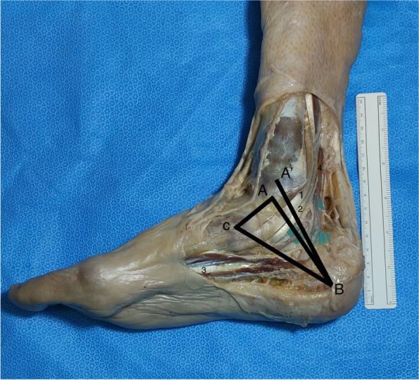
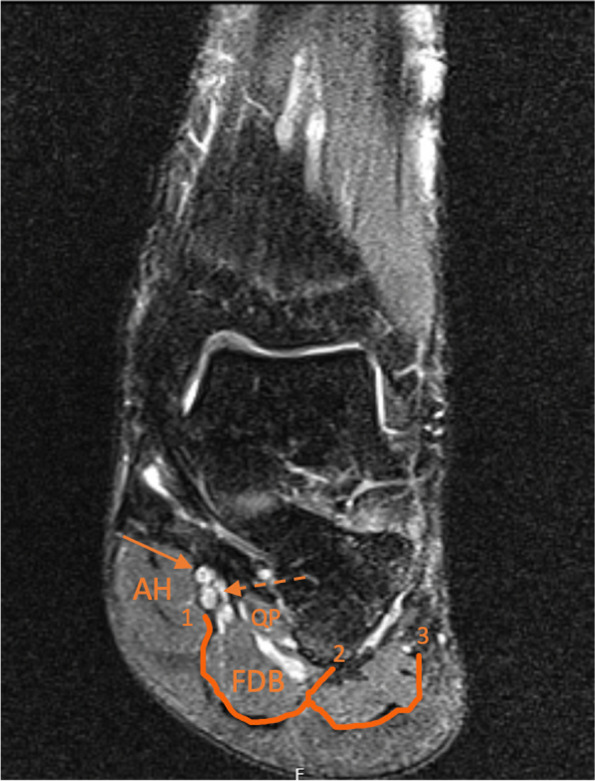
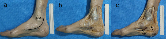
Results: The TN divided proximal to DML in 87.5%, on top of the DML in 12,5% and distal in none of the feet. The Baxter's nerve (BN) originated proximally in 50%, on top of the DML in 12,5% and distally in 37.5% of the cases. There was a strong and significant correlation between the length of DML and the distance from the center of the medial malleolus (MM) to the lateral plantar nerve (LPN), medial plantar (MPN) nerve, BN and Medial Calcaneal Nerve (MCN) (ρ: 0.910, 0.866, 0.970 and 0.762 respectively, p < 0.001).
Conclusions: In our sample the TN divides distal to DML in none of the cases. We also report a strong association between ankle size and the distribution of the MPN, LPN, BN and MCN. We hypothesize that location of these branches on the medial side of the ankle could be more predictable if we take into consideration the distance between the MM and the medial process of the calcaneal tuberosity.
期刊介绍:
Journal of Foot and Ankle Research, the official journal of the Australian Podiatry Association and The College of Podiatry (UK), is an open access journal that encompasses all aspects of policy, organisation, delivery and clinical practice related to the assessment, diagnosis, prevention and management of foot and ankle disorders.
Journal of Foot and Ankle Research covers a wide range of clinical subject areas, including diabetology, paediatrics, sports medicine, gerontology and geriatrics, foot surgery, physical therapy, dermatology, wound management, radiology, biomechanics and bioengineering, orthotics and prosthetics, as well the broad areas of epidemiology, policy, organisation and delivery of services related to foot and ankle care.
The journal encourages submissions from all health professionals who manage lower limb conditions, including podiatrists, nurses, physical therapists and physiotherapists, orthopaedists, manual therapists, medical specialists and general medical practitioners, as well as health service researchers concerned with foot and ankle care.
The Australian Podiatry Association and the College of Podiatry (UK) have reserve funds to cover the article-processing charge for manuscripts submitted by its members. Society members can email the appropriate contact at Australian Podiatry Association or The College of Podiatry to obtain the corresponding code to enter on submission.

 求助内容:
求助内容: 应助结果提醒方式:


