{"title":"Lymphatic vessels accompanying dorsal and basal dural sinuses in the human brain","authors":"Safiye Çavdar , Büşra Köse , Damlasu Altınöz , Gizem Söyler , Ahmet Cingöz , İlke Ali Gürses , Mazhar Özkan , Hızır Aslıyüksek , Halit Çakır","doi":"10.1016/j.jchemneu.2023.102357","DOIUrl":null,"url":null,"abstract":"<div><p>Recent investigations showed the presence of meningeal lymphatic vessels (mLVs) along the superior sagittal and transverse dural sinuses which drain both fluid and immune cells from the cerebrospinal fluid (CSF) to the deep cervical lymph nodes. This study uses immunohistochemistry (IHC) and the Western Blot technique to show the presence of mLV accompanying the dorsal (superior sagittal, inferior sagittal, transverse, sigmoid, and straight) and basal (cavernous, sphenoparietal, superior, and inferior petrosal) dural sinuses in the human brain. Samples for IHC were obtained from dorsal and basal meningeal dural sinuses of 3 human cadavers and 3 autopsies. Routine histological techniques were carried out for the specimens. Podoplanin (PDPN, lymphatic vessel endothelial cell marker) and CD31 (vascular endothelial cell marker) IHC staining were applied to the 5 µm thick paraffin sections. Furthermore, PDPN and CD31 protein expressions were evaluated using Western Blot to the tissue samples from the same regions of 4 autopsies. Two consecutive sections from each sinus were PDPN, and CD31 was stained to differentiate blood vessels (BV) from mLV. The IHC staining showed the presence of mLVs accompanying both dorsal and basal dural sinuses. The mLVs accompanying the dorsal dural sinuses had a larger dimensions range compared to the basal dural sinuses. However, the number of mLVs along the basal dural sinuses was more than the mLVs along the dorsal ones. Further, fluid channels were closely localized to the mLV, with varying diameters and densities. Western Blotting technique showed the presence of PDPN expression in both dorsal and basal dural sinus samples. The knowledge of the presence of mLV along both dorsal and basal dural sinuses in humans can increase the understanding of how mLV contributes to the brain lymphatic circulation and may help understand the neuropathophysiological processes of various neurological diseases.</p></div>","PeriodicalId":15324,"journal":{"name":"Journal of chemical neuroanatomy","volume":"134 ","pages":"Article 102357"},"PeriodicalIF":2.1000,"publicationDate":"2023-10-28","publicationTypes":"Journal Article","fieldsOfStudy":null,"isOpenAccess":false,"openAccessPdf":"","citationCount":"0","resultStr":null,"platform":"Semanticscholar","paperid":null,"PeriodicalName":"Journal of chemical neuroanatomy","FirstCategoryId":"3","ListUrlMain":"https://www.sciencedirect.com/science/article/pii/S0891061823001278","RegionNum":4,"RegionCategory":"医学","ArticlePicture":[],"TitleCN":null,"AbstractTextCN":null,"PMCID":null,"EPubDate":"","PubModel":"","JCR":"Q3","JCRName":"BIOCHEMISTRY & MOLECULAR BIOLOGY","Score":null,"Total":0}
引用次数: 0
Abstract
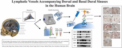
Recent investigations showed the presence of meningeal lymphatic vessels (mLVs) along the superior sagittal and transverse dural sinuses which drain both fluid and immune cells from the cerebrospinal fluid (CSF) to the deep cervical lymph nodes. This study uses immunohistochemistry (IHC) and the Western Blot technique to show the presence of mLV accompanying the dorsal (superior sagittal, inferior sagittal, transverse, sigmoid, and straight) and basal (cavernous, sphenoparietal, superior, and inferior petrosal) dural sinuses in the human brain. Samples for IHC were obtained from dorsal and basal meningeal dural sinuses of 3 human cadavers and 3 autopsies. Routine histological techniques were carried out for the specimens. Podoplanin (PDPN, lymphatic vessel endothelial cell marker) and CD31 (vascular endothelial cell marker) IHC staining were applied to the 5 µm thick paraffin sections. Furthermore, PDPN and CD31 protein expressions were evaluated using Western Blot to the tissue samples from the same regions of 4 autopsies. Two consecutive sections from each sinus were PDPN, and CD31 was stained to differentiate blood vessels (BV) from mLV. The IHC staining showed the presence of mLVs accompanying both dorsal and basal dural sinuses. The mLVs accompanying the dorsal dural sinuses had a larger dimensions range compared to the basal dural sinuses. However, the number of mLVs along the basal dural sinuses was more than the mLVs along the dorsal ones. Further, fluid channels were closely localized to the mLV, with varying diameters and densities. Western Blotting technique showed the presence of PDPN expression in both dorsal and basal dural sinus samples. The knowledge of the presence of mLV along both dorsal and basal dural sinuses in humans can increase the understanding of how mLV contributes to the brain lymphatic circulation and may help understand the neuropathophysiological processes of various neurological diseases.
期刊介绍:
The Journal of Chemical Neuroanatomy publishes scientific reports relating the functional and biochemical aspects of the nervous system with its microanatomical organization. The scope of the journal concentrates on reports which combine microanatomical, biochemical, pharmacological and behavioural approaches.
Papers should offer original data correlating the morphology of the nervous system (the brain and spinal cord in particular) with its biochemistry. The Journal of Chemical Neuroanatomy is particularly interested in publishing important studies performed with up-to-date methodology utilizing sensitive chemical microassays, hybridoma technology, immunocytochemistry, in situ hybridization and receptor radioautography, to name a few examples.
The Journal of Chemical Neuroanatomy is the natural vehicle for integrated studies utilizing these approaches. The articles will be selected by the editorial board and invited reviewers on the basis of their excellence and potential contribution to this field of neurosciences. Both in vivo and in vitro integrated studies in chemical neuroanatomy are appropriate subjects of interest to the journal. These studies should relate only to vertebrate species with particular emphasis on the mammalian and primate nervous systems.
 求助内容:
求助内容: 应助结果提醒方式:


