{"title":"A Successful Living Donor Liver Transplantation Using Hepatic Iron Deposition Graft Suspected by Magnetic Resonance Imaging.","authors":"Nobuhiko Kurata, Masato Shizuku, Kanta Jobara, Yoji Ishizu, Masatoshi Ishigami, Yasuhiro Ogura","doi":"10.1155/2023/9075184","DOIUrl":null,"url":null,"abstract":"<p><p>Recently, magnetic resonance imaging (MRI) has been developed as a widely available and noninvasive method for detecting and evaluating hepatic iron overload. This case report presents a successful living donor liver transplantation (LDLT) in which the donor was suspected to have hepatic iron deposition by MRI evaluation. A preoperative donor liver biopsy and genetic examination were performed to exclude hereditary hemochromatosis and other chronic liver diseases. A liver biopsy showed an almost normal liver specimen with a slight deposition of iron in 2-3% of hepatocytes, and a genetic examination of hereditary hemochromatosis revealed no typical mutations in HFE, TFR2, HJV, HAMP, or SLC40A1. Despite the traumatic hemothorax complication caused by the liver biopsy, the liver transplant eligibility was confirmed. Two months after the hemothorax complication, an LDLT donor operation was performed. The donor was discharged from the hospital on postoperative day (POD) #17 with favorable liver function. The recipient's posttransplant clinical course was generally favorable except for acute cellular rejection and biliary complications, and the recipient was discharged from the hospital on POD #87 with excellent graft function. A one-year follow-up liver biopsy of the recipient demonstrated almost normal liver with iron deposition in less than 1% of the hepatocytes, and no iron deposition was identified in the liver graft by MRI examination. Liver biopsy and genetic examination are effective methods to evaluate the eligibility of liver transplant donors with suspected hepatic iron deposition. The living donor with slight hepatic iron deposition, if hereditary hemochromatosis was ruled out, can donate partial liver safely.</p>","PeriodicalId":30327,"journal":{"name":"Case Reports in Transplantation","volume":"2023 ","pages":"9075184"},"PeriodicalIF":0.0000,"publicationDate":"2023-01-01","publicationTypes":"Journal Article","fieldsOfStudy":null,"isOpenAccess":false,"openAccessPdf":"https://www.ncbi.nlm.nih.gov/pmc/articles/PMC10036174/pdf/","citationCount":"0","resultStr":null,"platform":"Semanticscholar","paperid":null,"PeriodicalName":"Case Reports in Transplantation","FirstCategoryId":"1085","ListUrlMain":"https://doi.org/10.1155/2023/9075184","RegionNum":0,"RegionCategory":null,"ArticlePicture":[],"TitleCN":null,"AbstractTextCN":null,"PMCID":null,"EPubDate":"","PubModel":"","JCR":"","JCRName":"","Score":null,"Total":0}
引用次数: 0
Abstract
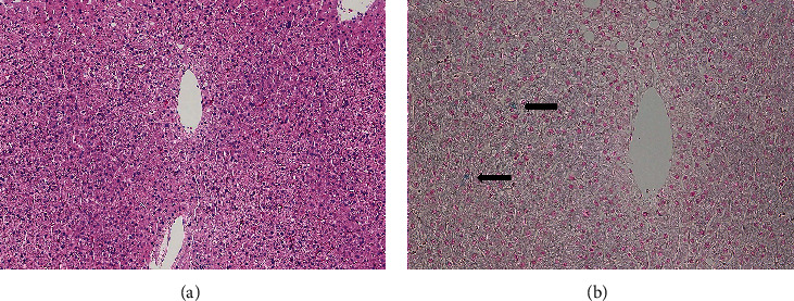
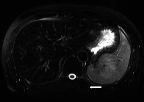
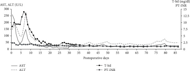
Recently, magnetic resonance imaging (MRI) has been developed as a widely available and noninvasive method for detecting and evaluating hepatic iron overload. This case report presents a successful living donor liver transplantation (LDLT) in which the donor was suspected to have hepatic iron deposition by MRI evaluation. A preoperative donor liver biopsy and genetic examination were performed to exclude hereditary hemochromatosis and other chronic liver diseases. A liver biopsy showed an almost normal liver specimen with a slight deposition of iron in 2-3% of hepatocytes, and a genetic examination of hereditary hemochromatosis revealed no typical mutations in HFE, TFR2, HJV, HAMP, or SLC40A1. Despite the traumatic hemothorax complication caused by the liver biopsy, the liver transplant eligibility was confirmed. Two months after the hemothorax complication, an LDLT donor operation was performed. The donor was discharged from the hospital on postoperative day (POD) #17 with favorable liver function. The recipient's posttransplant clinical course was generally favorable except for acute cellular rejection and biliary complications, and the recipient was discharged from the hospital on POD #87 with excellent graft function. A one-year follow-up liver biopsy of the recipient demonstrated almost normal liver with iron deposition in less than 1% of the hepatocytes, and no iron deposition was identified in the liver graft by MRI examination. Liver biopsy and genetic examination are effective methods to evaluate the eligibility of liver transplant donors with suspected hepatic iron deposition. The living donor with slight hepatic iron deposition, if hereditary hemochromatosis was ruled out, can donate partial liver safely.

 求助内容:
求助内容: 应助结果提醒方式:


