The correlation between different ultrasound planes and computed tomography measures of abdominal aortic aneurysms

Q3 Medicine
Brigid G Hill, Rossi Holloway, Joyce Lim, Kari Clifford, Sarah Lesche, James Letts, Jolanda Krysa
{"title":"The correlation between different ultrasound planes and computed tomography measures of abdominal aortic aneurysms","authors":"Brigid G Hill,&nbsp;Rossi Holloway,&nbsp;Joyce Lim,&nbsp;Kari Clifford,&nbsp;Sarah Lesche,&nbsp;James Letts,&nbsp;Jolanda Krysa","doi":"10.1002/ajum.12319","DOIUrl":null,"url":null,"abstract":"<div>\n \n \n <section>\n \n <h3> Introduction</h3>\n \n <p>Ultrasound measurements of the aorta are typically taken in the axial plane, with the transducer perpendicular to the aorta, and diameter measurements are obtained by placing the callipers from the anterior to the posterior wall and the transverse right to the left side of the aorta. While the ‘conventional’ anteroposterior walls in both sagittal and transverse plains may be suitable for aneurysms with less complicated geometry, there is controversy regarding the suitability of this approach for complicated, particularly tortuous aneurysms, as they may offer a more challenging situation. Previous work undertaken within our research group found that when training inexperienced users of ultrasound, they demonstrated more optimal calliper placement to the abdominal aorta when approached from a decubitus window to obtain a coronal image compared to the traditional ultrasound approach.</p>\n </section>\n \n <section>\n \n <h3> Purpose</h3>\n \n <p>To observe the level of agreement in real-world reporting between computed tomography (CT) and ultrasound measurements in three standard planes; transverse AP, sagittal AP and coronal (left to right) infra-renal abdominal aortic aneurysm (AAA) diameter.</p>\n </section>\n \n <section>\n \n <h3> Methodology</h3>\n \n <p>This is a retrospective review of the Otago Vascular Diagnostics database for AAA, where ultrasound and CT diameter data, available within 90 days of each other, were compared. In addition to patient demographics, the infrarenal aorta ultrasound diameter measurements in transverse AP and sagittal AP, along with a coronal decubitus image of the aorta was collected. No transverse measurement was performed from the left to the right of the aorta.</p>\n </section>\n \n <section>\n \n <h3> Results</h3>\n \n <p>Three hundred twenty-five participants (238 males, mean age 76.4 ± 7.5) were included. Mean ultrasound outer to the outer wall, transverse AP and sagittal AP diameters were 48.7 ± 10.5 mm and 48.9 ± 9.9 mm, respectively. The coronal diameter measurement of the aorta from left to right was 53.9 ± 12.8 mm in the left decubitus window. The mean ultrasound max was 54.3 ± 12.6 mm. The mean CT diameter measurement was 55.6 ± 12.7 mm. Correlation between the CT max and ultrasound max was <i>r</i><sup>2</sup> = 0.90, and CT with the coronal measurement <i>r</i><sup>2</sup> = 0.90, CT and AP transverse was r2=0.80, and CT with AP sagittal measurement was <i>r</i><sup>2</sup> = 0.77.</p>\n </section>\n \n <section>\n \n <h3> Conclusion</h3>\n \n <p>The decubitus ultrasound window of the abdominal aorta, with measurement of the coronal plane, is highly correlated and in agreement with CT scanning. This window may offer an alternative approach to measuring the infrarenal abdominal aortic aneurysm and should be considered when performing surveillance of all infra-renal AAA.</p>\n </section>\n </div>","PeriodicalId":36517,"journal":{"name":"Australasian Journal of Ultrasound in Medicine","volume":"26 1","pages":"5-12"},"PeriodicalIF":0.0000,"publicationDate":"2022-10-12","publicationTypes":"Journal Article","fieldsOfStudy":null,"isOpenAccess":false,"openAccessPdf":"https://onlinelibrary.wiley.com/doi/epdf/10.1002/ajum.12319","citationCount":"1","resultStr":null,"platform":"Semanticscholar","paperid":null,"PeriodicalName":"Australasian Journal of Ultrasound in Medicine","FirstCategoryId":"1085","ListUrlMain":"https://onlinelibrary.wiley.com/doi/10.1002/ajum.12319","RegionNum":0,"RegionCategory":null,"ArticlePicture":[],"TitleCN":null,"AbstractTextCN":null,"PMCID":null,"EPubDate":"","PubModel":"","JCR":"Q3","JCRName":"Medicine","Score":null,"Total":0}
引用次数: 1

Abstract

Introduction

Ultrasound measurements of the aorta are typically taken in the axial plane, with the transducer perpendicular to the aorta, and diameter measurements are obtained by placing the callipers from the anterior to the posterior wall and the transverse right to the left side of the aorta. While the ‘conventional’ anteroposterior walls in both sagittal and transverse plains may be suitable for aneurysms with less complicated geometry, there is controversy regarding the suitability of this approach for complicated, particularly tortuous aneurysms, as they may offer a more challenging situation. Previous work undertaken within our research group found that when training inexperienced users of ultrasound, they demonstrated more optimal calliper placement to the abdominal aorta when approached from a decubitus window to obtain a coronal image compared to the traditional ultrasound approach.

Purpose

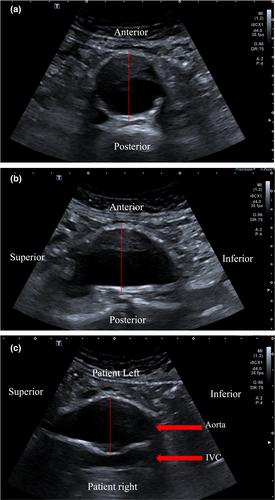
To observe the level of agreement in real-world reporting between computed tomography (CT) and ultrasound measurements in three standard planes; transverse AP, sagittal AP and coronal (left to right) infra-renal abdominal aortic aneurysm (AAA) diameter.

Methodology

This is a retrospective review of the Otago Vascular Diagnostics database for AAA, where ultrasound and CT diameter data, available within 90 days of each other, were compared. In addition to patient demographics, the infrarenal aorta ultrasound diameter measurements in transverse AP and sagittal AP, along with a coronal decubitus image of the aorta was collected. No transverse measurement was performed from the left to the right of the aorta.

Results

Three hundred twenty-five participants (238 males, mean age 76.4 ± 7.5) were included. Mean ultrasound outer to the outer wall, transverse AP and sagittal AP diameters were 48.7 ± 10.5 mm and 48.9 ± 9.9 mm, respectively. The coronal diameter measurement of the aorta from left to right was 53.9 ± 12.8 mm in the left decubitus window. The mean ultrasound max was 54.3 ± 12.6 mm. The mean CT diameter measurement was 55.6 ± 12.7 mm. Correlation between the CT max and ultrasound max was r2 = 0.90, and CT with the coronal measurement r2 = 0.90, CT and AP transverse was r2=0.80, and CT with AP sagittal measurement was r2 = 0.77.

Conclusion

The decubitus ultrasound window of the abdominal aorta, with measurement of the coronal plane, is highly correlated and in agreement with CT scanning. This window may offer an alternative approach to measuring the infrarenal abdominal aortic aneurysm and should be considered when performing surveillance of all infra-renal AAA.
Abstract Image

不同超声平面与腹主动脉瘤计算机断层扫描测量之间的相关性。
简介:主动脉的超声测量通常在轴向平面内进行,换能器垂直于主动脉,直径测量是通过从主动脉前壁到后壁和从右到左横向放置卡尺来获得的。虽然矢状面和横向平面的“传统”前后壁可能适用于几何形状不太复杂的动脉瘤,但对于这种方法是否适用于复杂的,特别是弯曲的动脉瘤存在争议,因为它们可能会带来更具挑战性的情况。我们研究小组先前的工作发现,在培训缺乏经验的超声用户时,与传统的超声方法相比,他们在从卧位窗口接近腹主动脉以获得冠状图像时,表现出了更优化的卡尺位置。目的:观察计算机断层扫描(CT)和超声测量在三个标准平面上的真实世界报告的一致性水平;横向AP、矢状AP和冠状(从左到右)肾下腹主动脉瘤(AAA)直径。方法:这是对AAA的Otago血管诊断数据库的回顾性审查,其中超声和CT直径数据在90内可用 相互的天数进行比较。除了患者的人口统计数据外,还收集了肾下主动脉横向AP和矢状AP的超声直径测量值,以及主动脉的冠状卧位图像。从主动脉的左侧到右侧没有进行横向测量。结果:325名参与者(238名男性,平均年龄76.4岁) ± 7.5)。平均超声外径至外壁、横向AP和矢状AP直径为48.7 ± 10.5 mm和48.9 ± 9.9 mm。主动脉的冠状直径测量值从左到右为53.9 ± 12.8 mm在左侧卧位窗口。平均超声最大值为54.3 ± 12.6 平均CT直径测量值为55.6 ± 12.7 CT最大值与超声最大值的相关性为r2 = 0.90,CT与冠状测量r2 = 0.90,CT和AP横向测量r2=0.80,CT与AP矢状测量r 2 = 0.77.结论:腹主动脉卧位超声窗与冠状面测量高度相关,与CT扫描一致。该窗口可能为测量肾下腹主动脉瘤提供了一种替代方法,在监测所有肾下AAA时应予以考虑。
本文章由计算机程序翻译,如有差异,请以英文原文为准。
求助全文
约1分钟内获得全文 求助全文
来源期刊
Australasian Journal of Ultrasound in Medicine
Australasian Journal of Ultrasound in Medicine Medicine-Radiology, Nuclear Medicine and Imaging
CiteScore
1.90
自引率
0.00%
发文量
40
×
引用
GB/T 7714-2015
复制
MLA
复制
APA
复制
导出至
BibTeX EndNote RefMan NoteFirst NoteExpress
×
提示
您的信息不完整,为了账户安全,请先补充。
现在去补充
×
提示
您因"违规操作"
具体请查看互助需知
我知道了
×
提示
确定
请完成安全验证×
copy
已复制链接
快去分享给好友吧!
我知道了
右上角分享
点击右上角分享
0
联系我们:info@booksci.cn Book学术提供免费学术资源搜索服务,方便国内外学者检索中英文文献。致力于提供最便捷和优质的服务体验。 Copyright © 2023 布克学术 All rights reserved.
京ICP备2023020795号-1
ghs 京公网安备 11010802042870号
Book学术文献互助
Book学术文献互助群
群 号:481959085
Book学术官方微信