Propolis anti-inflammatory effects on MAGE-1 and retinoic acid-treated dendritic cells and on Th1 and T regulatory cells.
IF 1.8
3区 医学
Q4 TOXICOLOGY
Journal of Venomous Animals and Toxins Including Tropical Diseases
Pub Date : 2023-01-01
DOI:10.1590/1678-9199-JVATITD-2022-0044
引用次数: 1
Abstract
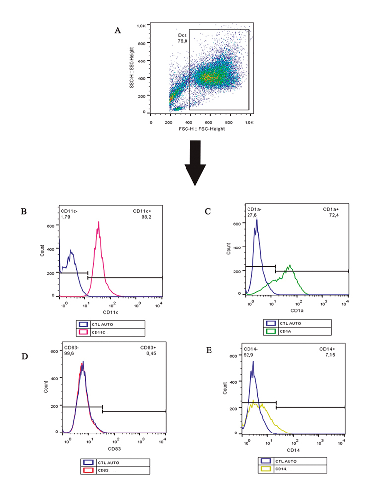
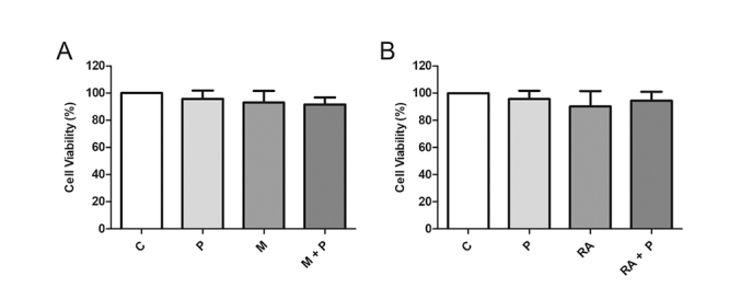
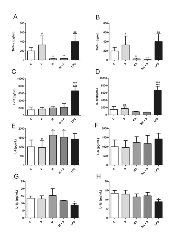
Abstract Background: Propolis exhibits huge potential in the pharmaceutical industry. In the present study, its effects were investigated on dendritic cells (DCs) stimulated with a tumor antigen (MAGE-1) and retinoic acid (RA) and on T lymphocytes to observe a possible differential activation of T lymphocytes, driving preferentially to Th1 or Treg cells. Methods: Cell viability, lymphocyte proliferation, gene expression (T-bet and FoxP3), and cytokine production by DCs (TNF-α, IL-10, IL-6 and IL-1β) and lymphocytes (IFN-γ and TGF-β) were analyzed. Results: MAGE-1 and RA alone or in combination with propolis inhibited TNF-α production and induced a higher lymphoproliferation compared to control, while MAGE-1 + propolis induced IL-6 production. Propolis in combination with RA induced FoxP3 expression. MAGE-1 induced IFN-γ production while propolis inhibited it, returning to basal levels. RA inhibited TGF-β production, what was counteracted by propolis. Conclusion: Propolis affected immunological parameters inhibiting pro-inflammatory cytokines and favoring the regulatory profile, opening perspectives for the control of inflammatory conditions.
蜂胶对MAGE-1和维甲酸处理的树突状细胞以及Th1和T调节细胞的抗炎作用。
背景:蜂胶在制药行业显示出巨大的潜力。在本研究中,研究了其对肿瘤抗原(MAGE-1)和视黄酸(RA)刺激的树突状细胞(DCs)和T淋巴细胞的影响,以观察T淋巴细胞可能的差异活化,优先驱动Th1或Treg细胞。方法:分析细胞活力、淋巴细胞增殖、基因表达(T-bet、FoxP3)及dc (TNF-α、IL-10、IL-6、IL-1β)和淋巴细胞(IFN-γ、TGF-β)细胞因子的产生。结果:与对照组相比,MAGE-1和RA单独或联合蜂胶抑制TNF-α的产生,诱导更高的淋巴细胞增殖,而MAGE-1 +蜂胶诱导IL-6的产生。蜂胶联合RA诱导FoxP3表达。MAGE-1诱导IFN-γ产生,蜂胶抑制IFN-γ产生,使其恢复到基础水平。RA抑制TGF-β的产生,蜂胶抵消了这一作用。结论:蜂胶影响免疫参数,抑制促炎细胞因子,有利于炎症的调控,为炎症的控制开辟了新的思路。
本文章由计算机程序翻译,如有差异,请以英文原文为准。
求助全文
约1分钟内获得全文
求助全文
来源期刊
CiteScore
4.80
自引率
8.30%
发文量
39
审稿时长
6-12 weeks
期刊介绍:
Journal of Venomous Animals and Toxins including Tropical Diseases (JVATiTD) is a non-commercial academic open access publication dedicated to research on all aspects of toxinology, venomous animals and tropical diseases. Its interdisciplinary content includes original scientific articles covering research on toxins derived from animals, plants and microorganisms. Topics of interest include, but are not limited to:systematics and morphology of venomous animals;physiology, biochemistry, pharmacology and immunology of toxins;epidemiology, clinical aspects and treatment of envenoming by different animals, plants and microorganisms;development and evaluation of antivenoms and toxin-derivative products;epidemiology, clinical aspects and treatment of tropical diseases (caused by virus, bacteria, algae, fungi and parasites) including the neglected tropical diseases (NTDs) defined by the World Health Organization.
 求助内容:
求助内容: 应助结果提醒方式:


