Ashleigh Barrett-Young, Wickliffe C Abraham, Carol Y Cheung, Jesse Gale, Sean Hogan, David Ireland, Ross Keenan, Annchen R Knodt, Tracy R Melzer, Terrie E Moffitt, Sandhya Ramrakha, Yih Chung Tham, Graham A Wilson, Tien Yin Wong, Ahmad R Hariri, Richie Poulton
{"title":"Associations Between Thinner Retinal Neuronal Layers and Suboptimal Brain Structural Integrity in a Middle-Aged Cohort.","authors":"Ashleigh Barrett-Young, Wickliffe C Abraham, Carol Y Cheung, Jesse Gale, Sean Hogan, David Ireland, Ross Keenan, Annchen R Knodt, Tracy R Melzer, Terrie E Moffitt, Sandhya Ramrakha, Yih Chung Tham, Graham A Wilson, Tien Yin Wong, Ahmad R Hariri, Richie Poulton","doi":"10.2147/EB.S402510","DOIUrl":null,"url":null,"abstract":"<p><strong>Purpose: </strong>The retina has potential as a biomarker of brain health and Alzheimer's disease (AD) because it is the only part of the central nervous system which can be easily imaged and has advantages over brain imaging technologies. Few studies have compared retinal and brain measurements in a middle-aged sample. The objective of our study was to investigate whether retinal neuronal measurements were associated with structural brain measurements in a middle-aged population-based cohort.</p><p><strong>Participants and methods: </strong>Participants were members of the Dunedin Multidisciplinary Health and Development Study (n=1037; a longitudinal cohort followed from birth and at ages 3, 5, 7, 9, 11, 13, 15, 18, 21, 26, 32, 38, and most recently at age 45, when 94% of the living Study members participated). Retinal nerve fibre layer (RNFL) and ganglion cell-inner plexiform layer (GC-IPL) thickness were measured by optical coherence tomography (OCT). Brain age gap estimate (brainAGE), cortical surface area, cortical thickness, subcortical grey matter volumes, white matter hyperintensities, were measured by magnetic resonance imaging (MRI).</p><p><strong>Results: </strong>Participants with both MRI and OCT data were included in the analysis (RNFL n=828, female n=413 [49.9%], male n=415 [50.1%]; GC-IPL n=825, female n=413 [50.1%], male n=412 [49.9%]). Thinner retinal neuronal layers were associated with older brain age, smaller cortical surface area, thinner average cortex, smaller subcortical grey matter volumes, and increased volume of white matter hyperintensities.</p><p><strong>Conclusion: </strong>These findings provide evidence that the retinal neuronal layers reflect differences in midlife structural brain integrity consistent with increased risk for later AD, supporting the proposition that the retina may be an early biomarker of brain health.</p>","PeriodicalId":51844,"journal":{"name":"Eye and Brain","volume":"15 ","pages":"25-35"},"PeriodicalIF":2.4000,"publicationDate":"2023-03-11","publicationTypes":"Journal Article","fieldsOfStudy":null,"isOpenAccess":false,"openAccessPdf":"https://ftp.ncbi.nlm.nih.gov/pub/pmc/oa_pdf/1e/7b/eb-15-25.PMC10018220.pdf","citationCount":"0","resultStr":null,"platform":"Semanticscholar","paperid":null,"PeriodicalName":"Eye and Brain","FirstCategoryId":"1085","ListUrlMain":"https://doi.org/10.2147/EB.S402510","RegionNum":0,"RegionCategory":null,"ArticlePicture":[],"TitleCN":null,"AbstractTextCN":null,"PMCID":null,"EPubDate":"2023/1/1 0:00:00","PubModel":"eCollection","JCR":"Q1","JCRName":"OPHTHALMOLOGY","Score":null,"Total":0}
引用次数: 0
Abstract
Purpose: The retina has potential as a biomarker of brain health and Alzheimer's disease (AD) because it is the only part of the central nervous system which can be easily imaged and has advantages over brain imaging technologies. Few studies have compared retinal and brain measurements in a middle-aged sample. The objective of our study was to investigate whether retinal neuronal measurements were associated with structural brain measurements in a middle-aged population-based cohort.
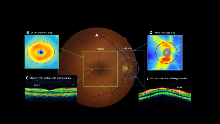
Participants and methods: Participants were members of the Dunedin Multidisciplinary Health and Development Study (n=1037; a longitudinal cohort followed from birth and at ages 3, 5, 7, 9, 11, 13, 15, 18, 21, 26, 32, 38, and most recently at age 45, when 94% of the living Study members participated). Retinal nerve fibre layer (RNFL) and ganglion cell-inner plexiform layer (GC-IPL) thickness were measured by optical coherence tomography (OCT). Brain age gap estimate (brainAGE), cortical surface area, cortical thickness, subcortical grey matter volumes, white matter hyperintensities, were measured by magnetic resonance imaging (MRI).
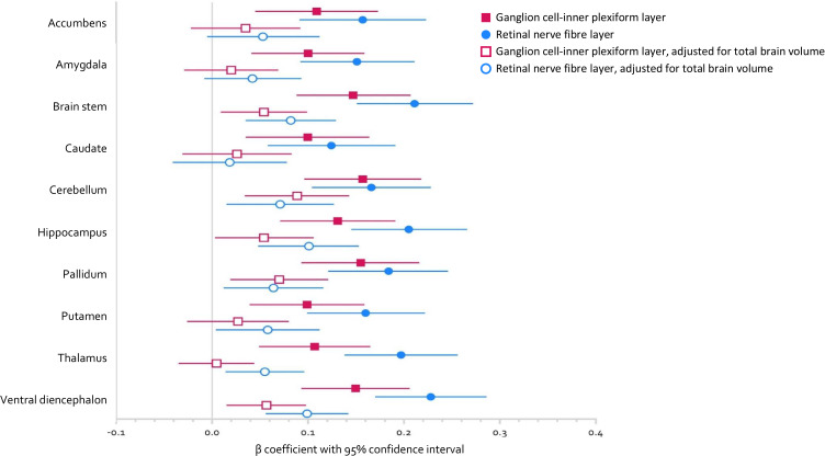
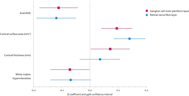
Results: Participants with both MRI and OCT data were included in the analysis (RNFL n=828, female n=413 [49.9%], male n=415 [50.1%]; GC-IPL n=825, female n=413 [50.1%], male n=412 [49.9%]). Thinner retinal neuronal layers were associated with older brain age, smaller cortical surface area, thinner average cortex, smaller subcortical grey matter volumes, and increased volume of white matter hyperintensities.
Conclusion: These findings provide evidence that the retinal neuronal layers reflect differences in midlife structural brain integrity consistent with increased risk for later AD, supporting the proposition that the retina may be an early biomarker of brain health.
期刊介绍:
Eye and Brain is an international, peer-reviewed, open access journal focusing on basic research, clinical findings, and expert reviews in the field of visual science and neuro-ophthalmology. The journal’s unique focus is the link between two well-known visual centres, the eye and the brain, with an emphasis on the importance of such connections. All aspects of clinical and especially basic research on the visual system are addressed within the journal as well as significant future directions in vision research and therapeutic measures. This unique journal focuses on neurological aspects of vision – both physiological and pathological. The scope of the journal spans from the cornea to the associational visual cortex and all the visual centers in between. Topics range from basic biological mechanisms to therapeutic treatment, from simple organisms to humans, and utilizing techniques from molecular biology to behavior. The journal especially welcomes primary research articles or review papers that make the connection between the eye and the brain. Specific areas covered in the journal include: Physiology and pathophysiology of visual centers, Eye movement disorders and strabismus, Cellular, biochemical, and molecular features of the visual system, Structural and functional organization of the eye and of the visual cortex, Metabolic demands of the visual system, Diseases and disorders with neuro-ophthalmic manifestations, Clinical and experimental neuro-ophthalmology and visual system pathologies, Epidemiological studies.

 求助内容:
求助内容: 应助结果提醒方式:


