Gemstone Spectral CT Virtual Noncontrast Images and Iodine Maps for the Characterization of Thyroid Lesions and Distinguishing Thyroid Papillary Carcinoma from Nodular Goiter.
Chun Yao, Xiaofeng Chen, Zhiqi Yang, Ruibin Huang, Sheng Zhang, Yuting Liao, Xiangguang Chen, Zhuozhi Dai
{"title":"Gemstone Spectral CT Virtual Noncontrast Images and Iodine Maps for the Characterization of Thyroid Lesions and Distinguishing Thyroid Papillary Carcinoma from Nodular Goiter.","authors":"Chun Yao, Xiaofeng Chen, Zhiqi Yang, Ruibin Huang, Sheng Zhang, Yuting Liao, Xiangguang Chen, Zhuozhi Dai","doi":"10.1155/2023/8220034","DOIUrl":null,"url":null,"abstract":"<p><strong>Background: </strong>Gemstone spectral contrast-enhanced CT with virtual noncontrast (VNC) images and iodine maps can potentially reduce the number of required CT scans for thyroid lesions. However, data regarding the clinical utility of VNC images and iodine maps in characterizing thyroid lesions and distinguishing thyroid papillary carcinoma from nodular goiter are still limited.</p><p><strong>Purpose: </strong>To determine whether VNC images and iodine density could reliably aid in characterizing thyroid lesions and distinguishing thyroid papillary carcinoma from nodular goiter compared with true noncontrast (TNC) images.</p><p><strong>Methods: </strong>This retrospective study included patients with thyroid papillary carcinoma or nodular goiter who underwent TNC and contrast-enhanced gemstone spectral CT scans. The consistency of qualitative parameters, including intralesional calcification, necrosis, lesion boundary, thyroid edge interruption, and lymph node metastasis, between TNC and VNC images, was analyzed using the kappa statistic. TNC attenuation, VNC attenuation, absolute attenuation between TNC and VNC, and iodine density were compared between thyroid papillary carcinoma and nodular goiter by using Student's <i>t</i>-test. The diagnostic performance for distinguishing papillary carcinoma from nodular goiter was evaluated by using the area under the receiver operating characteristic curve (AUC) value, sensitivity, and specificity.</p><p><strong>Results: </strong>VNC and TNC imaging showed comparable performance in delineating calcification, necrosis, lesion boundary, thyroid edge interruption, and lymph node metastasis (all <i>k</i> > 0.75). Papillary carcinoma showed significantly lower absolute attenuation between VNC and TNC than nodular goiter (7.86 ± 6.74 vs. 13.43 ± 10.53, <i>P</i>=0.026), which was similarly observed for iodine density (31.45 ± 8.51 vs. 37.27 ± 10.34, <i>P</i>=0.016). The iodine density showed higher diagnostic performance (AUC = 0.727), accuracy (0.773 vs. 0.667), sensitivity (0.750 vs. 0.708), and specificity (0.786 vs. 0.643) than the absolute attenuation between TNC and VNC images (AUC = 0.683).</p><p><strong>Conclusions: </strong>VNC imaging, a promising substitute for TNC imaging, has comparable diagnostic efficacy for reliably characterizing thyroid lesions. Iodine density could be valuable for distinguishing thyroid papillary carcinoma from nodular goiter.</p>","PeriodicalId":13966,"journal":{"name":"International Journal of Endocrinology","volume":"2023 ","pages":"8220034"},"PeriodicalIF":2.3000,"publicationDate":"2023-01-01","publicationTypes":"Journal Article","fieldsOfStudy":null,"isOpenAccess":false,"openAccessPdf":"https://www.ncbi.nlm.nih.gov/pmc/articles/PMC9988381/pdf/","citationCount":"0","resultStr":null,"platform":"Semanticscholar","paperid":null,"PeriodicalName":"International Journal of Endocrinology","FirstCategoryId":"3","ListUrlMain":"https://doi.org/10.1155/2023/8220034","RegionNum":4,"RegionCategory":"医学","ArticlePicture":[],"TitleCN":null,"AbstractTextCN":null,"PMCID":null,"EPubDate":"","PubModel":"","JCR":"Q3","JCRName":"ENDOCRINOLOGY & METABOLISM","Score":null,"Total":0}
引用次数: 0
Abstract
Background: Gemstone spectral contrast-enhanced CT with virtual noncontrast (VNC) images and iodine maps can potentially reduce the number of required CT scans for thyroid lesions. However, data regarding the clinical utility of VNC images and iodine maps in characterizing thyroid lesions and distinguishing thyroid papillary carcinoma from nodular goiter are still limited.
Purpose: To determine whether VNC images and iodine density could reliably aid in characterizing thyroid lesions and distinguishing thyroid papillary carcinoma from nodular goiter compared with true noncontrast (TNC) images.
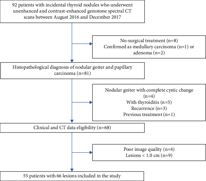
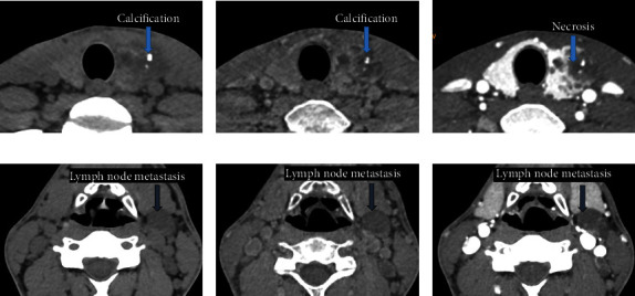
Methods: This retrospective study included patients with thyroid papillary carcinoma or nodular goiter who underwent TNC and contrast-enhanced gemstone spectral CT scans. The consistency of qualitative parameters, including intralesional calcification, necrosis, lesion boundary, thyroid edge interruption, and lymph node metastasis, between TNC and VNC images, was analyzed using the kappa statistic. TNC attenuation, VNC attenuation, absolute attenuation between TNC and VNC, and iodine density were compared between thyroid papillary carcinoma and nodular goiter by using Student's t-test. The diagnostic performance for distinguishing papillary carcinoma from nodular goiter was evaluated by using the area under the receiver operating characteristic curve (AUC) value, sensitivity, and specificity.
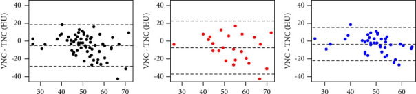
Results: VNC and TNC imaging showed comparable performance in delineating calcification, necrosis, lesion boundary, thyroid edge interruption, and lymph node metastasis (all k > 0.75). Papillary carcinoma showed significantly lower absolute attenuation between VNC and TNC than nodular goiter (7.86 ± 6.74 vs. 13.43 ± 10.53, P=0.026), which was similarly observed for iodine density (31.45 ± 8.51 vs. 37.27 ± 10.34, P=0.016). The iodine density showed higher diagnostic performance (AUC = 0.727), accuracy (0.773 vs. 0.667), sensitivity (0.750 vs. 0.708), and specificity (0.786 vs. 0.643) than the absolute attenuation between TNC and VNC images (AUC = 0.683).
Conclusions: VNC imaging, a promising substitute for TNC imaging, has comparable diagnostic efficacy for reliably characterizing thyroid lesions. Iodine density could be valuable for distinguishing thyroid papillary carcinoma from nodular goiter.
背景:宝石光谱对比增强CT与虚拟非对比(VNC)图像和碘图可以潜在地减少所需的甲状腺病变的CT扫描次数。然而,关于VNC图像和碘图在诊断甲状腺病变和区分甲状腺乳头状癌和结节性甲状腺肿方面的临床应用的数据仍然有限。目的:确定VNC图像和碘密度是否能可靠地帮助诊断甲状腺病变和区分甲状腺乳头状癌与结节性甲状腺肿。方法:本回顾性研究纳入了接受TNC和增强宝石光谱CT扫描的甲状腺乳头状癌或结节性甲状腺肿患者。采用kappa统计分析TNC与VNC影像间病灶内钙化、坏死、病灶边界、甲状腺边缘中断、淋巴结转移等定性参数的一致性。采用Student’st检验比较甲状腺乳头状癌和结节性甲状腺肿的TNC衰减、VNC衰减、TNC与VNC之间的绝对衰减以及碘密度。通过使用受者工作特征曲线下面积(AUC)值、敏感性和特异性来评估鉴别乳头状癌和结节性甲状腺肿的诊断性能。结果:VNC和TNC成像在描绘钙化、坏死、病变边界、甲状腺边缘中断和淋巴结转移方面表现相当(k均> 0.75)。乳头状癌在VNC和TNC间的绝对衰减明显低于结节性甲状腺肿(7.86±6.74比13.43±10.53,P=0.026),碘密度的绝对衰减与乳头状癌相似(31.45±8.51比37.27±10.34,P=0.016)。碘密度的诊断效能(AUC = 0.727)、准确性(0.773 vs. 0.667)、敏感性(0.750 vs. 0.708)和特异性(0.786 vs. 0.643)均高于TNC和VNC影像间的绝对衰减(AUC = 0.683)。结论:VNC成像是一种有希望的替代TNC成像的方法,在可靠地诊断甲状腺病变方面具有相当的疗效。碘密度对鉴别甲状腺乳头状癌和结节性甲状腺肿有价值。
期刊介绍:
International Journal of Endocrinology is a peer-reviewed, Open Access journal that provides a forum for scientists and clinicians working in basic and translational research. The journal publishes original research articles, review articles, and clinical studies that provide insights into the endocrine system and its associated diseases at a genomic, molecular, biochemical and cellular level.
 求助内容:
求助内容: 应助结果提醒方式:


