{"title":"Survival Rate and Cervical Bone Loss of Dental Implants Placed in Regenerated Areas with Free Iliac Graft.","authors":"Gholamreza Shirani, Mahboubeh Hashemi Nasab, Shahryar Bashiri, Sheida Kordi","doi":"10.30476/DENTJODS.2022.90184.1474","DOIUrl":null,"url":null,"abstract":"<p><strong>Statement of the problem: </strong>For many years, practitioners have been encountered with dental rehabilitation of atrophic jaws. Among many of alternatives, free iliac graft can be a reasonable and also problematic choice to be accomplished.</p><p><strong>Purpose: </strong>The aim of this study was to evaluate the implant survival rate and bone loss in implants inserted in reconstructed jaws with free iliac graft.</p><p><strong>Materials and method: </strong>In this clinical trial study, twelve patients that underwent bone reconstruction with free iliac graft were included in this retrospective study. The patients underwent surgery over a 6-year period from September 2011 to July 2017. Panoramic images were taken immediately after implant insertion and at the follow-up session. The parameters that were assessed included implant survival rate, bone level changes, and surrounding tissue conditions.</p><p><strong>Results: </strong>One hundred and nine implants were placed in eight female and four male patients, of which 65 (59.6%) were inserted in the reconstructed maxilla and 44 (40.3%) in the reconstructed mandible. The interval between the reconstruction surgery and follow-up session was 28.75 months and the mean interval between implant insertion and the follow-up session was 21.75 months, ranging from 6 to 72 months. The total average of crestal bone resorption was 2.44 mm (range: 0 to 5.43 mm).</p><p><strong>Conclusion: </strong>This study found that rehabilitation of atrophic jaws with dental implants placed in free iliac graft was associated with acceptable marginal bone loss, survival rate, satisfaction, and esthetic results among the patients.</p>","PeriodicalId":73702,"journal":{"name":"Journal of dentistry (Shiraz, Iran)","volume":"24 1","pages":"53-59"},"PeriodicalIF":0.0000,"publicationDate":"2023-03-01","publicationTypes":"Journal Article","fieldsOfStudy":null,"isOpenAccess":false,"openAccessPdf":"https://www.ncbi.nlm.nih.gov/pmc/articles/PMC9971605/pdf/","citationCount":"0","resultStr":null,"platform":"Semanticscholar","paperid":null,"PeriodicalName":"Journal of dentistry (Shiraz, Iran)","FirstCategoryId":"1085","ListUrlMain":"https://doi.org/10.30476/DENTJODS.2022.90184.1474","RegionNum":0,"RegionCategory":null,"ArticlePicture":[],"TitleCN":null,"AbstractTextCN":null,"PMCID":null,"EPubDate":"","PubModel":"","JCR":"","JCRName":"","Score":null,"Total":0}
引用次数: 0
Abstract
Statement of the problem: For many years, practitioners have been encountered with dental rehabilitation of atrophic jaws. Among many of alternatives, free iliac graft can be a reasonable and also problematic choice to be accomplished.
Purpose: The aim of this study was to evaluate the implant survival rate and bone loss in implants inserted in reconstructed jaws with free iliac graft.
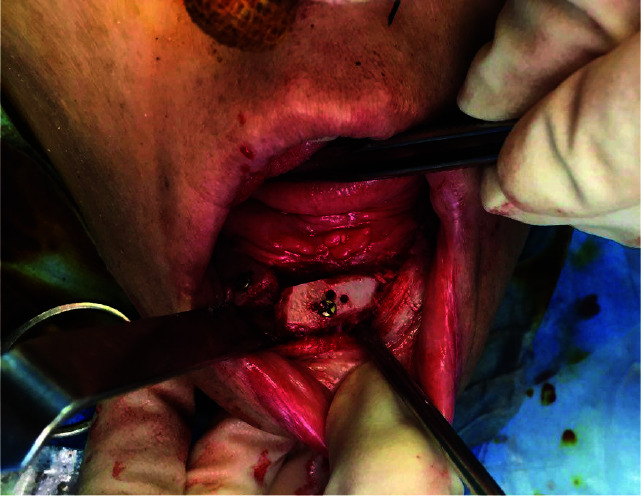
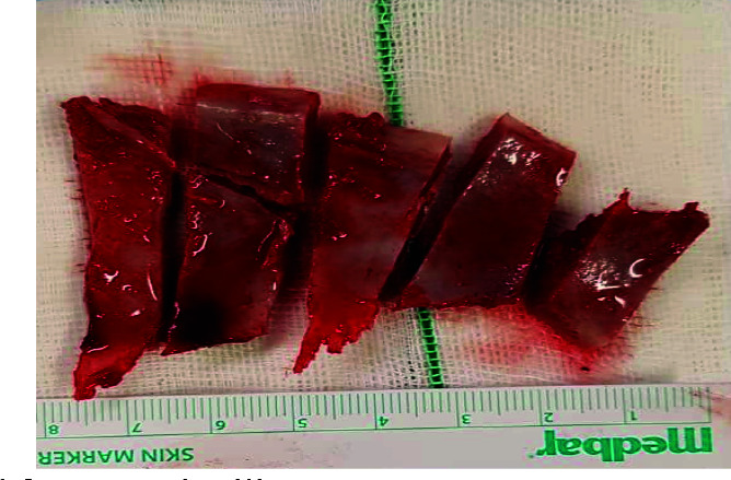
Materials and method: In this clinical trial study, twelve patients that underwent bone reconstruction with free iliac graft were included in this retrospective study. The patients underwent surgery over a 6-year period from September 2011 to July 2017. Panoramic images were taken immediately after implant insertion and at the follow-up session. The parameters that were assessed included implant survival rate, bone level changes, and surrounding tissue conditions.
Results: One hundred and nine implants were placed in eight female and four male patients, of which 65 (59.6%) were inserted in the reconstructed maxilla and 44 (40.3%) in the reconstructed mandible. The interval between the reconstruction surgery and follow-up session was 28.75 months and the mean interval between implant insertion and the follow-up session was 21.75 months, ranging from 6 to 72 months. The total average of crestal bone resorption was 2.44 mm (range: 0 to 5.43 mm).
Conclusion: This study found that rehabilitation of atrophic jaws with dental implants placed in free iliac graft was associated with acceptable marginal bone loss, survival rate, satisfaction, and esthetic results among the patients.


 求助内容:
求助内容: 应助结果提醒方式:


