{"title":"Stress Induction and Release: Electroencephalography Study on Brain Networks and Cortisol.","authors":"Zahra Rezvani, Reza Khosrowabadi, Afrooz Seyedebrahimi, Golam-Hossein Meftahi, Boshra Hatef","doi":"10.32598/bcn.2021.2525.1","DOIUrl":null,"url":null,"abstract":"<p><strong>Introduction: </strong>Acute stress over a long time period can drastically influence the behavioral and cognitive performances. Therefore, it is important to control and eliminate the stressor after a stressful event. In this regard, understanding of brain mechanism of stress release will help to introduce new practical approaches. In this study, we aimed to investigate the changes in the brain's functional connectivity (FC) patterns and salivary cortisol level during stress induction and release in healthy young male adults.</p><p><strong>Method: </strong>In this study, 20 healthy young male adults were exposed to stressful events using the Trier social stress paradigm in one session consisting of 23 minutes of psychological stress induction and 20 minutes of recovery, Their stress was measured by the visual analog scale (VAS). In addition, their salivary cortisol levels and electroencephalography (EEG) data were recorded. Subsequently, brain FC maps were prepared in a frequency-specific manner. Then, the effects of inducing and releasing stress on the VAS, cortisol level, and FC were assessed.</p><p><strong>Results: </strong>The inter-hemispheric FC of the right frontal lobes with other brain regions decreased, while the FC was increased in the left frontal lobes during the induction of stress. Interestingly, the release of stress presented a recovery pattern of inter-hemispheric FC. These changes in FC significantly correlated with changes in the cortisol level.</p><p><strong>Conclusion: </strong>Our findings highlight the important role of bihemispheric associations in adaptation and coping with stressful conditions.</p>","PeriodicalId":8728,"journal":{"name":"Basic and Clinical Neuroscience Journal","volume":"4 1","pages":"607-616"},"PeriodicalIF":0.0000,"publicationDate":"2024-09-01","publicationTypes":"Journal Article","fieldsOfStudy":null,"isOpenAccess":false,"openAccessPdf":"https://www.ncbi.nlm.nih.gov/pmc/articles/PMC12198744/pdf/","citationCount":"0","resultStr":null,"platform":"Semanticscholar","paperid":null,"PeriodicalName":"Basic and Clinical Neuroscience Journal","FirstCategoryId":"1085","ListUrlMain":"https://doi.org/10.32598/bcn.2021.2525.1","RegionNum":0,"RegionCategory":null,"ArticlePicture":[],"TitleCN":null,"AbstractTextCN":null,"PMCID":null,"EPubDate":"","PubModel":"","JCR":"","JCRName":"","Score":null,"Total":0}
引用次数: 0
Abstract
Introduction: Acute stress over a long time period can drastically influence the behavioral and cognitive performances. Therefore, it is important to control and eliminate the stressor after a stressful event. In this regard, understanding of brain mechanism of stress release will help to introduce new practical approaches. In this study, we aimed to investigate the changes in the brain's functional connectivity (FC) patterns and salivary cortisol level during stress induction and release in healthy young male adults.
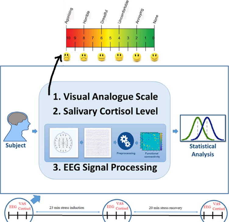
Method: In this study, 20 healthy young male adults were exposed to stressful events using the Trier social stress paradigm in one session consisting of 23 minutes of psychological stress induction and 20 minutes of recovery, Their stress was measured by the visual analog scale (VAS). In addition, their salivary cortisol levels and electroencephalography (EEG) data were recorded. Subsequently, brain FC maps were prepared in a frequency-specific manner. Then, the effects of inducing and releasing stress on the VAS, cortisol level, and FC were assessed.
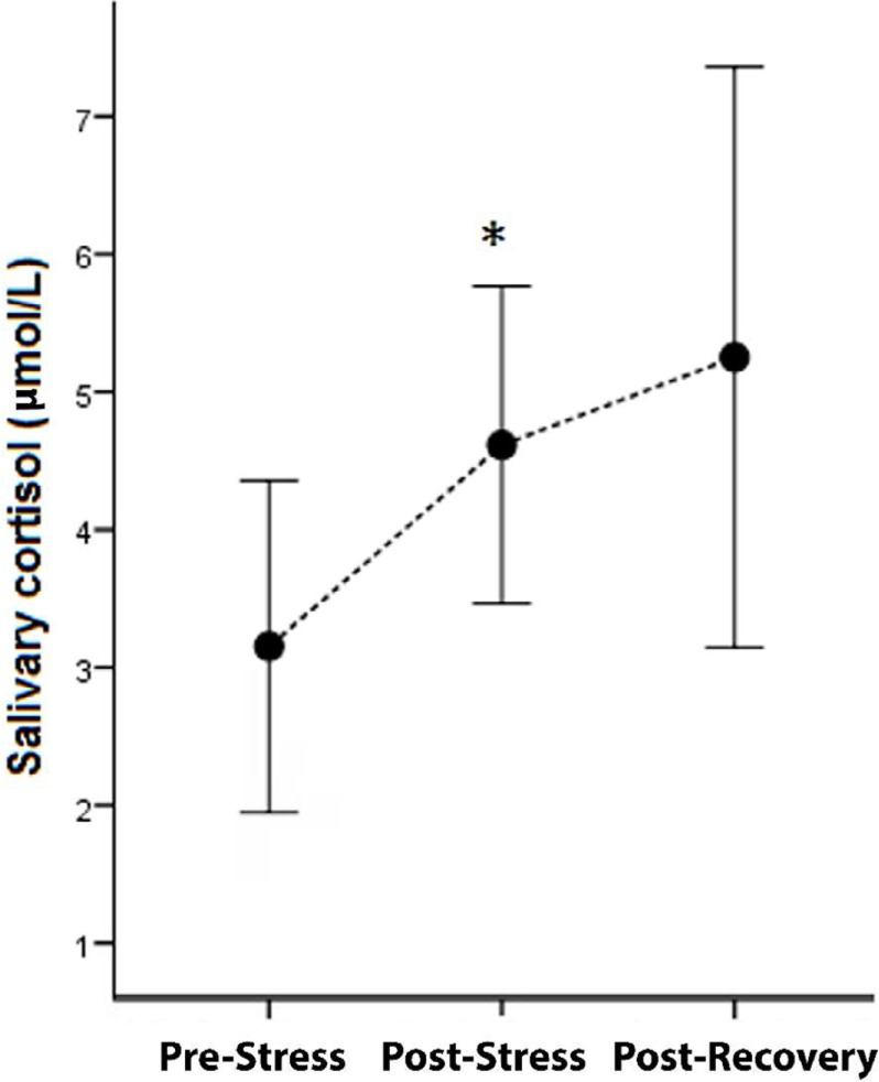
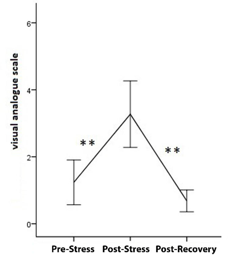
Results: The inter-hemispheric FC of the right frontal lobes with other brain regions decreased, while the FC was increased in the left frontal lobes during the induction of stress. Interestingly, the release of stress presented a recovery pattern of inter-hemispheric FC. These changes in FC significantly correlated with changes in the cortisol level.
Conclusion: Our findings highlight the important role of bihemispheric associations in adaptation and coping with stressful conditions.

 求助内容:
求助内容: 应助结果提醒方式:


