{"title":"Near-infrared-II-activated photothermal nanotransducers for wireless neuronal stimulation","authors":"Xianzhe Tang, Zhaowei Chen, Huangyao Yang","doi":"10.1002/mba2.15","DOIUrl":null,"url":null,"abstract":"<p>Recently, Wu et al.<span><sup>1</sup></span> presented an interesting study using near-infrared II (NIR-II)-activated photothermal nanotransducers for remote deep-brain stimulation (DBS) in freely behaving animals in an efficient and safe fashion. This study provided a complementary method for state-of-the-art technologies utilized for DBS. DBS with superior spatial-temporal precision would hold great promise for clinical management of brain disorders and fundamental neuroscience and offer unique advantages compared to brain lesioning procedures regarding reversibility and adaptability.<span><sup>2</sup></span></p><p>Over the past decades, a host of strategies have been developed for the modulation of neurons deep in the brain.<span><sup>3</sup></span> To name a few, conventional electrical stimulation with implantable microelectrodes has been widely applied for DBS, which, however, suffers from coarse temporal resolution and chronic immune responses (e.g., gliosis) at the implantation site of brain tissues.<span><sup>2</sup></span> As a technology showing the revolutionary impact on neurobiology, optogenetics holds great potential in elucidation or manipulation of specific neurons and neural circuits with precise timings and locations.<span><sup>4</sup></span> In this paradigm, to minimize the scattering of light in the brain, invasive optical fibers must be inserted to deliver photons to the target neurons which are infected with opsin-expressing vectors. The implantation of optical fibers easily causes permanent damage to the brain tissues and physically perturbates animals' natural movement, confining conventional optogenetics to limited applications.<span><sup>5</sup></span> Recent advances in sonogenetics, sono-optogenetics, and magnetothermal genetics have allowed the dissection of neuron circuits via implant-free and tether-free stimulation strategies.<span><sup>3</sup></span> Nevertheless, limitation remains for these technologies because the activity sphere for animal behavior manipulation is spatially confined around a resonant coil or a focused ultrasound beam.<span><sup>3</sup></span></p><p>Alternatively, NIR (700–1700 nm in wavelength) light has emerged for tether-less deep-brain modulation with the assistance of upconversion and photothermal micro- and nanoparticles as the transducers.<span><sup>6-8</sup></span> The 808 nm laser, a common NIR-I (700–900 nm) illumination source, has been leveraged for modulating neural activity with Nd-doped upconversion nanoparticles. Attributing to the low absorption coefficient of water at such a wavelength, the overheating side effect caused by NIR irradiation was mitigated, yet its limited penetration depth (1–2 mm) hindered its application for DBS. Meanwhile, although 980 nm NIR-II excited Yb-doped upconversion transducers have shown certain promises in modulating deep brain neurons, there are still concerns associated with nonspecific tissue heating. Therefore, further improvements are desired to pursuit novel DBS method that can obviate implantation, tether-fibers, limitation of activity arenas, light attenuation, and thermal biodamage in brain tissues. Now, writing in <i>Nature Biomedical Engineering</i>, Wu et al.<span><sup>1</sup></span> reported an implant- and tether-free 1064 nm NIR-II photothermal DBS in free-behaving mice with deeper brain penetration and wider-field illumination using polymeric nanoparticle transducers (Figure 1A).</p><p>To realize their design, Wu et al.<span><sup>1</sup></span> first synthesized NIR-II excited photothermal nanomaterials with semiconducting poly(benzobisthiadiazole-alt-vinylene) as the core and poly(lactide-co-glycolide)-<i>b</i>-poly(ethylene glycol) as a shell, by which they called macromolecular infrared nanotransducers for deep-brain stimulation (MINDS). The photothermal conversion efficiency of MINDS was measured to be 71% under 1064 nm irradiation at 10 mW mm<sup>−2</sup>. An important reason why they choseD this excitation wavelength was because it had rather low brain tissue attenuation.<span><sup>1</sup></span> In addition, to engage neurons with MINDS under NIR-II irradiation, transient receptor potential cation channel subfamily V member 1 (TRPV1), a kind of temperature-sensitive nonselective cation channels, were ectopically expressed in neurons located in specific brain regions and then elicited neurons to fire action potentials in response to NIR-II irradiation-triggered local temperature increase (Figure 1B).</p><p>To validate the feasibility of photothermal neuronal modulation with the assistance of MINDS, TRPV1 channels were transduced in HEK293T cells in vitro followed by low-intensity NIR-II illumination and dynamic calcium imaging. The temporal variation in the calcium signal in HEK293T cells with different treatments demonstrated the selective 1064 nm NIR-II activation of MINDS-sensitized TRPV1 channels at the cellular level, validating the reliability of the NIR-II photothermal genetic toolkit.</p><p>TRPV1 adeno-associated viral vector and MINDS were sequentially injected into the hippocampus or the secondary motor cortex (M2) of the mouse brain, then a distant 1064 nm illumination light was placed above the head. In vivo electrophysiological recording, immunohistological staining, and tether-free unilateral circling behavioral tests within a wide arena were then carried out (Figure 1C). Increase in neuron firing rate, c-Fos expression, and rotation speed of mice collectively revealed that TRPV1, MINDS, and NIR-II irradiation could be well engaged to activate the neuron cells in the M2 region. Notably, a distant power density of 8 mW mm<sup>−2</sup>, within the safe limit for the 1064 nm illumination, increased ∼2°C at the target brain region. Benefiting from the deep penetration of 1064 nm NIR-II illumination and the superior photothermal performance of MINDS, such a low power density was still sufficient to activate TRPV1 and endow in vivo NIR-II photothermal neuromodulation with a short on- and offset response time in a wide arena for motor behavioral study.</p><p>Taking advantage of the deep penetration of 1064 nm light through the scalp and skull, Wu et al.<span><sup>1</sup></span> further utilized the NIR-II photothermal genetic toolkit to modulate neural activity in the ventral tegmental area (VTA) in deep-brain regions and control the reward circuitry-associated conditioned place preference behavior. After sequentially tagging dopaminergic neurons by using TRPV1-encoding adeno-associated viral vectors and injecting MINDS there, contextual conditioning test with NIR-II neuromodulation was carried out in a Y maze (Figure 1D). Successive conditioned place preference results revealed that mice with both TRPV1 overexpression and MINDS injection in VTA spent a longer time in the NIR-II illuminated arm terminal. Despite the deep location of VTA inside the brain, the “biological transparent” character of 1064 nm NIR-II illumination made it easy to get the desired temperature (i.e., ∼39.1°C) in the VTA region merely by adjusting the incident power density. More importantly, this implant- and tether-free paradigm allowed free motion and interaction of mice by placing the 1064 nm light sources ∼1 m above animals' head. Of note, this study revealed that, at a power density of 10 mW mm<sup>−2</sup>, thermal damage to the brain is insignificant.</p><p>Overall, the study by Wu et al.<span><sup>1</sup></span> presented a pioneering work showing 1064 nm NIR-II neural stimulation by combining excellent photothermal nanotransducers with temperature-sensitive TRPV1. With this NIR-II photothermal genetic toolkit, they modulated specific neuronal activities in multiple brain regions of varying depths and controlled mouse behaviors in wide arenas.</p><p>Considering that NIR-II-triggered temperature changes in other brain regions may influence the physiological activity, future full-brain mapping of the optimal light- and nanotransducer-delivery parameters would be necessary for further applications.<span><sup>9</sup></span> Regarding the thicker skin and skull compared to that of rodents, the millimeter-range penetration depth of 1064 nm irradiation remains a stumbling block for optogenetic DBS on nonhuman primates and human subjects. Transition of this toolkit from laboratory to clinical practice will require further optimization of the dose of transducers and the power of laser for matching the complexity of human brains. The combination of this photothermal DBS method with devices and probes for imaging and sensing neural dynamics may help to improve the accuracy of manipulating specific neurons and neural circuits.<span><sup>10</sup></span> In parallel, inspired by the success of engineering opsin,<span><sup>9</sup></span> systematic optimization of TRPV1 channels for higher sensitivity will allow for lower temperature enhancement and shorter response time for photothermal DBS. These advances would make it possible to achieve more precise and efficient neural circuits dissection and neurological disorder therapy.</p><p>Xianzhe Tang and Zhaowei Chen drafted the manuscript. Huangyao Yang revised the manuscript.</p><p>The authors declare no conflict of interest.</p><p>The ethics statement is not applicable for this study.</p>","PeriodicalId":100901,"journal":{"name":"MedComm – Biomaterials and Applications","volume":"1 2","pages":""},"PeriodicalIF":0.0000,"publicationDate":"2022-09-23","publicationTypes":"Journal Article","fieldsOfStudy":null,"isOpenAccess":false,"openAccessPdf":"https://onlinelibrary.wiley.com/doi/epdf/10.1002/mba2.15","citationCount":"0","resultStr":null,"platform":"Semanticscholar","paperid":null,"PeriodicalName":"MedComm – Biomaterials and Applications","FirstCategoryId":"1085","ListUrlMain":"https://onlinelibrary.wiley.com/doi/10.1002/mba2.15","RegionNum":0,"RegionCategory":null,"ArticlePicture":[],"TitleCN":null,"AbstractTextCN":null,"PMCID":null,"EPubDate":"","PubModel":"","JCR":"","JCRName":"","Score":null,"Total":0}
引用次数: 0
Abstract
Recently, Wu et al.1 presented an interesting study using near-infrared II (NIR-II)-activated photothermal nanotransducers for remote deep-brain stimulation (DBS) in freely behaving animals in an efficient and safe fashion. This study provided a complementary method for state-of-the-art technologies utilized for DBS. DBS with superior spatial-temporal precision would hold great promise for clinical management of brain disorders and fundamental neuroscience and offer unique advantages compared to brain lesioning procedures regarding reversibility and adaptability.2
Over the past decades, a host of strategies have been developed for the modulation of neurons deep in the brain.3 To name a few, conventional electrical stimulation with implantable microelectrodes has been widely applied for DBS, which, however, suffers from coarse temporal resolution and chronic immune responses (e.g., gliosis) at the implantation site of brain tissues.2 As a technology showing the revolutionary impact on neurobiology, optogenetics holds great potential in elucidation or manipulation of specific neurons and neural circuits with precise timings and locations.4 In this paradigm, to minimize the scattering of light in the brain, invasive optical fibers must be inserted to deliver photons to the target neurons which are infected with opsin-expressing vectors. The implantation of optical fibers easily causes permanent damage to the brain tissues and physically perturbates animals' natural movement, confining conventional optogenetics to limited applications.5 Recent advances in sonogenetics, sono-optogenetics, and magnetothermal genetics have allowed the dissection of neuron circuits via implant-free and tether-free stimulation strategies.3 Nevertheless, limitation remains for these technologies because the activity sphere for animal behavior manipulation is spatially confined around a resonant coil or a focused ultrasound beam.3
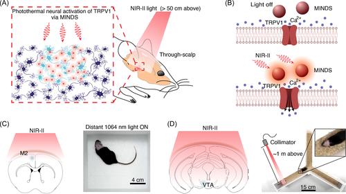
Alternatively, NIR (700–1700 nm in wavelength) light has emerged for tether-less deep-brain modulation with the assistance of upconversion and photothermal micro- and nanoparticles as the transducers.6-8 The 808 nm laser, a common NIR-I (700–900 nm) illumination source, has been leveraged for modulating neural activity with Nd-doped upconversion nanoparticles. Attributing to the low absorption coefficient of water at such a wavelength, the overheating side effect caused by NIR irradiation was mitigated, yet its limited penetration depth (1–2 mm) hindered its application for DBS. Meanwhile, although 980 nm NIR-II excited Yb-doped upconversion transducers have shown certain promises in modulating deep brain neurons, there are still concerns associated with nonspecific tissue heating. Therefore, further improvements are desired to pursuit novel DBS method that can obviate implantation, tether-fibers, limitation of activity arenas, light attenuation, and thermal biodamage in brain tissues. Now, writing in Nature Biomedical Engineering, Wu et al.1 reported an implant- and tether-free 1064 nm NIR-II photothermal DBS in free-behaving mice with deeper brain penetration and wider-field illumination using polymeric nanoparticle transducers (Figure 1A).
To realize their design, Wu et al.1 first synthesized NIR-II excited photothermal nanomaterials with semiconducting poly(benzobisthiadiazole-alt-vinylene) as the core and poly(lactide-co-glycolide)-b-poly(ethylene glycol) as a shell, by which they called macromolecular infrared nanotransducers for deep-brain stimulation (MINDS). The photothermal conversion efficiency of MINDS was measured to be 71% under 1064 nm irradiation at 10 mW mm−2. An important reason why they choseD this excitation wavelength was because it had rather low brain tissue attenuation.1 In addition, to engage neurons with MINDS under NIR-II irradiation, transient receptor potential cation channel subfamily V member 1 (TRPV1), a kind of temperature-sensitive nonselective cation channels, were ectopically expressed in neurons located in specific brain regions and then elicited neurons to fire action potentials in response to NIR-II irradiation-triggered local temperature increase (Figure 1B).
To validate the feasibility of photothermal neuronal modulation with the assistance of MINDS, TRPV1 channels were transduced in HEK293T cells in vitro followed by low-intensity NIR-II illumination and dynamic calcium imaging. The temporal variation in the calcium signal in HEK293T cells with different treatments demonstrated the selective 1064 nm NIR-II activation of MINDS-sensitized TRPV1 channels at the cellular level, validating the reliability of the NIR-II photothermal genetic toolkit.
TRPV1 adeno-associated viral vector and MINDS were sequentially injected into the hippocampus or the secondary motor cortex (M2) of the mouse brain, then a distant 1064 nm illumination light was placed above the head. In vivo electrophysiological recording, immunohistological staining, and tether-free unilateral circling behavioral tests within a wide arena were then carried out (Figure 1C). Increase in neuron firing rate, c-Fos expression, and rotation speed of mice collectively revealed that TRPV1, MINDS, and NIR-II irradiation could be well engaged to activate the neuron cells in the M2 region. Notably, a distant power density of 8 mW mm−2, within the safe limit for the 1064 nm illumination, increased ∼2°C at the target brain region. Benefiting from the deep penetration of 1064 nm NIR-II illumination and the superior photothermal performance of MINDS, such a low power density was still sufficient to activate TRPV1 and endow in vivo NIR-II photothermal neuromodulation with a short on- and offset response time in a wide arena for motor behavioral study.
Taking advantage of the deep penetration of 1064 nm light through the scalp and skull, Wu et al.1 further utilized the NIR-II photothermal genetic toolkit to modulate neural activity in the ventral tegmental area (VTA) in deep-brain regions and control the reward circuitry-associated conditioned place preference behavior. After sequentially tagging dopaminergic neurons by using TRPV1-encoding adeno-associated viral vectors and injecting MINDS there, contextual conditioning test with NIR-II neuromodulation was carried out in a Y maze (Figure 1D). Successive conditioned place preference results revealed that mice with both TRPV1 overexpression and MINDS injection in VTA spent a longer time in the NIR-II illuminated arm terminal. Despite the deep location of VTA inside the brain, the “biological transparent” character of 1064 nm NIR-II illumination made it easy to get the desired temperature (i.e., ∼39.1°C) in the VTA region merely by adjusting the incident power density. More importantly, this implant- and tether-free paradigm allowed free motion and interaction of mice by placing the 1064 nm light sources ∼1 m above animals' head. Of note, this study revealed that, at a power density of 10 mW mm−2, thermal damage to the brain is insignificant.
Overall, the study by Wu et al.1 presented a pioneering work showing 1064 nm NIR-II neural stimulation by combining excellent photothermal nanotransducers with temperature-sensitive TRPV1. With this NIR-II photothermal genetic toolkit, they modulated specific neuronal activities in multiple brain regions of varying depths and controlled mouse behaviors in wide arenas.
Considering that NIR-II-triggered temperature changes in other brain regions may influence the physiological activity, future full-brain mapping of the optimal light- and nanotransducer-delivery parameters would be necessary for further applications.9 Regarding the thicker skin and skull compared to that of rodents, the millimeter-range penetration depth of 1064 nm irradiation remains a stumbling block for optogenetic DBS on nonhuman primates and human subjects. Transition of this toolkit from laboratory to clinical practice will require further optimization of the dose of transducers and the power of laser for matching the complexity of human brains. The combination of this photothermal DBS method with devices and probes for imaging and sensing neural dynamics may help to improve the accuracy of manipulating specific neurons and neural circuits.10 In parallel, inspired by the success of engineering opsin,9 systematic optimization of TRPV1 channels for higher sensitivity will allow for lower temperature enhancement and shorter response time for photothermal DBS. These advances would make it possible to achieve more precise and efficient neural circuits dissection and neurological disorder therapy.
Xianzhe Tang and Zhaowei Chen drafted the manuscript. Huangyao Yang revised the manuscript.
The authors declare no conflict of interest.
The ethics statement is not applicable for this study.

 求助内容:
求助内容: 应助结果提醒方式:


