A classification of the plantar intrinsic foot muscles based on the physiological cross-sectional area and muscle fiber length in healthy young adult males.
{"title":"A classification of the plantar intrinsic foot muscles based on the physiological cross-sectional area and muscle fiber length in healthy young adult males.","authors":"Yuki Kusagawa, Toshiyuki Kurihara, Sumiaki Maeo, Takashi Sugiyama, Hiroaki Kanehisa, Tadao Isaka","doi":"10.1186/s13047-023-00676-2","DOIUrl":null,"url":null,"abstract":"<p><strong>Background: </strong>Plantar intrinsic foot muscles (PIFMs) are composed of 10 muscles and play an essential role in achieving functional diversity in the foot. Previous studies have identified that the morphological profiles of PIFMs vary between individuals. The morphological profiles of a muscle theoretically reflect its output potentials: the physiological cross-sectional area (PCSA) of a muscle is proportional to its maximum force generation, and the muscle fiber length (FL) is its shortening velocity. This implies that the PCSA and FL may be useful variables for characterizing the functional diversity of the individual PIFM. The purpose of this study was to examine how individual PIFMs can be classified based on their PCSA and FL.</p><p><strong>Methods: </strong>In 26 healthy young adult males, the muscle volume and muscle length of seven PIFMs (abductor hallucis, ABDH; abductor digiti minimi, ABDM; adductor hallucis oblique head, ADDH-OH; ADDH transverse head, ADDH-TH; flexor digitorum brevis, FDB; flexor hallucis brevis, FHB; quadratus plantae, QP) were measured using magnetic resonance imaging. The PCSA and FL of each of the seven PIFMs were then estimated by combining the data measured from the participants and those of muscle architectural parameters documented from cadavers in previous studies. A total of 182 data samples (26 participants × 7 muscles) were classified into clusters using k-means cluster analysis. The optimal number of clusters was evaluated using the elbow method.</p><p><strong>Results: </strong>The data samples of PIFMs were assigned to four clusters with different morphological profiles: ADDH-OH and FHB, characterised by large PCSA and short FL (high force generation and slow shortening velocity potentials); ABDM and FDB, moderate PCSA and moderate FL (moderate force generation and moderate shortening velocity potentials); QP, moderate PCSA and long FL (moderate force generation and rapid shortening velocity potentials); ADDH-TH, small PCSA and moderate FL (low force generation and moderate shortening velocity potentials). ABDH components were assigned equivalently to the first and second clusters.</p><p><strong>Conclusions: </strong>The approach adopted in this study may provide a novel perspective for interpreting the PIFMs' function based on their maximal force generation and shortening velocity potentials.</p>","PeriodicalId":49164,"journal":{"name":"Journal of Foot and Ankle Research","volume":"16 1","pages":"75"},"PeriodicalIF":2.2000,"publicationDate":"2023-11-11","publicationTypes":"Journal Article","fieldsOfStudy":null,"isOpenAccess":false,"openAccessPdf":"https://www.ncbi.nlm.nih.gov/pmc/articles/PMC10638735/pdf/","citationCount":"0","resultStr":null,"platform":"Semanticscholar","paperid":null,"PeriodicalName":"Journal of Foot and Ankle Research","FirstCategoryId":"3","ListUrlMain":"https://doi.org/10.1186/s13047-023-00676-2","RegionNum":3,"RegionCategory":"医学","ArticlePicture":[],"TitleCN":null,"AbstractTextCN":null,"PMCID":null,"EPubDate":"","PubModel":"","JCR":"Q1","JCRName":"ORTHOPEDICS","Score":null,"Total":0}
引用次数: 0
Abstract
Background: Plantar intrinsic foot muscles (PIFMs) are composed of 10 muscles and play an essential role in achieving functional diversity in the foot. Previous studies have identified that the morphological profiles of PIFMs vary between individuals. The morphological profiles of a muscle theoretically reflect its output potentials: the physiological cross-sectional area (PCSA) of a muscle is proportional to its maximum force generation, and the muscle fiber length (FL) is its shortening velocity. This implies that the PCSA and FL may be useful variables for characterizing the functional diversity of the individual PIFM. The purpose of this study was to examine how individual PIFMs can be classified based on their PCSA and FL.
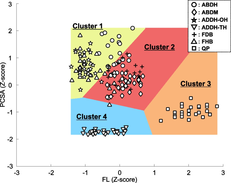
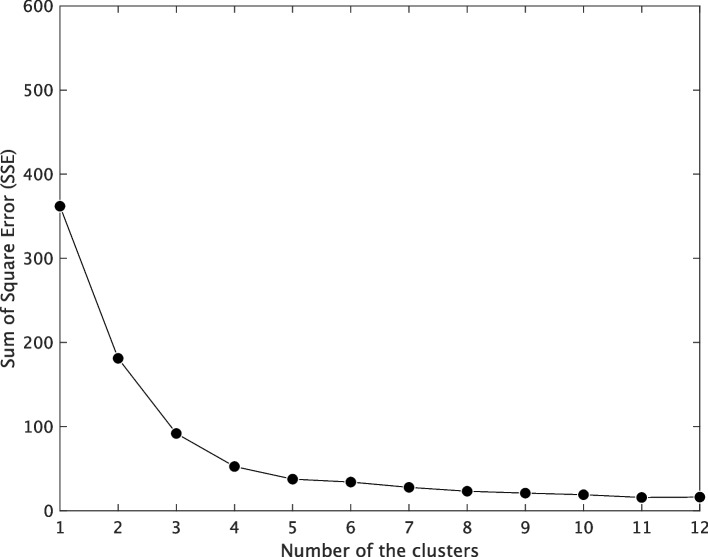
Methods: In 26 healthy young adult males, the muscle volume and muscle length of seven PIFMs (abductor hallucis, ABDH; abductor digiti minimi, ABDM; adductor hallucis oblique head, ADDH-OH; ADDH transverse head, ADDH-TH; flexor digitorum brevis, FDB; flexor hallucis brevis, FHB; quadratus plantae, QP) were measured using magnetic resonance imaging. The PCSA and FL of each of the seven PIFMs were then estimated by combining the data measured from the participants and those of muscle architectural parameters documented from cadavers in previous studies. A total of 182 data samples (26 participants × 7 muscles) were classified into clusters using k-means cluster analysis. The optimal number of clusters was evaluated using the elbow method.
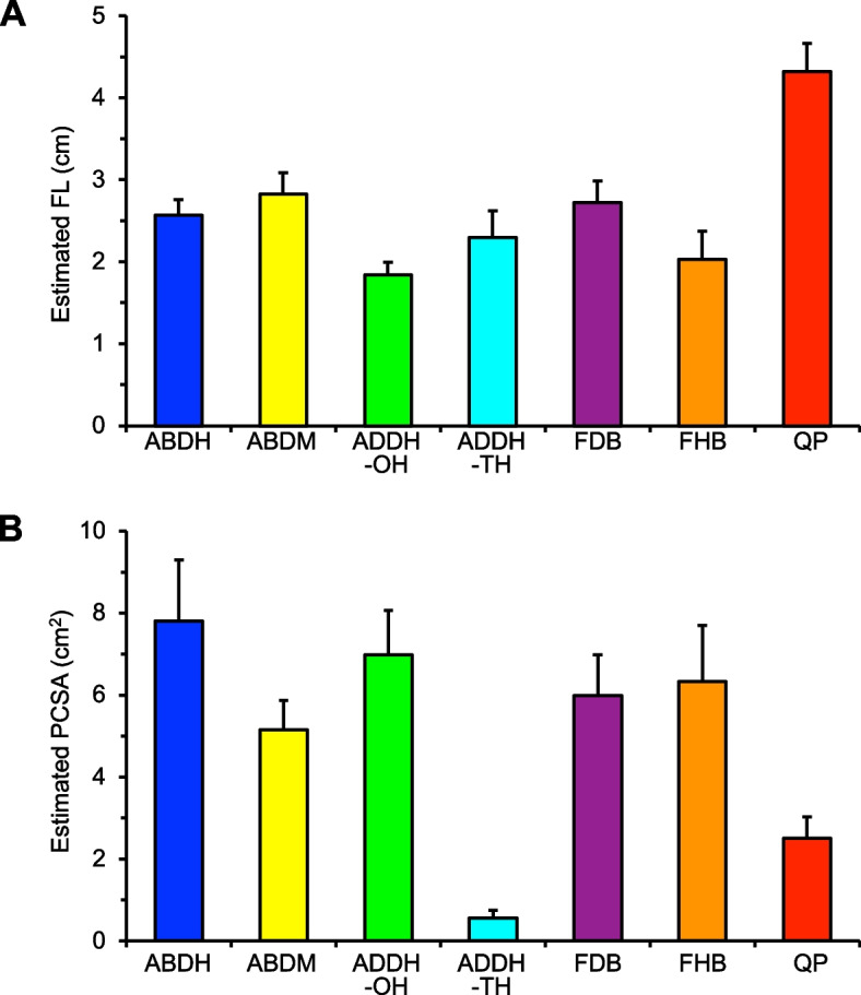
Results: The data samples of PIFMs were assigned to four clusters with different morphological profiles: ADDH-OH and FHB, characterised by large PCSA and short FL (high force generation and slow shortening velocity potentials); ABDM and FDB, moderate PCSA and moderate FL (moderate force generation and moderate shortening velocity potentials); QP, moderate PCSA and long FL (moderate force generation and rapid shortening velocity potentials); ADDH-TH, small PCSA and moderate FL (low force generation and moderate shortening velocity potentials). ABDH components were assigned equivalently to the first and second clusters.
Conclusions: The approach adopted in this study may provide a novel perspective for interpreting the PIFMs' function based on their maximal force generation and shortening velocity potentials.
期刊介绍:
Journal of Foot and Ankle Research, the official journal of the Australian Podiatry Association and The College of Podiatry (UK), is an open access journal that encompasses all aspects of policy, organisation, delivery and clinical practice related to the assessment, diagnosis, prevention and management of foot and ankle disorders.
Journal of Foot and Ankle Research covers a wide range of clinical subject areas, including diabetology, paediatrics, sports medicine, gerontology and geriatrics, foot surgery, physical therapy, dermatology, wound management, radiology, biomechanics and bioengineering, orthotics and prosthetics, as well the broad areas of epidemiology, policy, organisation and delivery of services related to foot and ankle care.
The journal encourages submissions from all health professionals who manage lower limb conditions, including podiatrists, nurses, physical therapists and physiotherapists, orthopaedists, manual therapists, medical specialists and general medical practitioners, as well as health service researchers concerned with foot and ankle care.
The Australian Podiatry Association and the College of Podiatry (UK) have reserve funds to cover the article-processing charge for manuscripts submitted by its members. Society members can email the appropriate contact at Australian Podiatry Association or The College of Podiatry to obtain the corresponding code to enter on submission.
 求助内容:
求助内容: 应助结果提醒方式:


