Harsimran Kaur, Parakriti Gupta, Haseen Ahmad, Shamanth A Shankarnarayan, Sonakshi Srivastava, Suneeta Sahu, T Karuna, Tarun Narang, Sunita Gupta, Anup Ghosh, Shivaprakash M Rudramurthy
{"title":"Cladosporium halotolerans: Exploring an Unheeded Human Pathogen.","authors":"Harsimran Kaur, Parakriti Gupta, Haseen Ahmad, Shamanth A Shankarnarayan, Sonakshi Srivastava, Suneeta Sahu, T Karuna, Tarun Narang, Sunita Gupta, Anup Ghosh, Shivaprakash M Rudramurthy","doi":"10.1007/s11046-023-00801-6","DOIUrl":null,"url":null,"abstract":"<p><strong>Background: </strong>Cladosporium halotolerans is a saprobic fungus, rarely implicated in human infections. The identification is challenging due to non-specific phenotypic features.</p><p><strong>Objective: </strong>To decipher clinical spectrum, microbiological and susceptibility profile of clinical and environmental isolates of Cladosporium halotolerans.</p><p><strong>Method: </strong>All the isolates identified as Cladosporium halotolerans deposited in National Culture Collection for Pathogenic Fungi (NCCPF), Postgraduate Institute of Medical Education and Research, Chandigarh, India were revived. Phenotypic and molecular characterization targeting internal transcribed spacer (ITS) region of ribosomal DNA, large subunit of ribosomal DNA (LSU; NL1 and NL4), actin (ACT) and beta-tubulin (TUB) was done. Scanning electron microscopy (SEM) was performed to determine any phenotypic variations. Antifungal susceptibility testing (AFST) was carried out for eight antifungal agents as per CLSI M38 Ed3 guidelines. We also performed systematic literature review of all the cases of Cladosporium halotolerans reported till date.</p><p><strong>Results: </strong>A total of four isolates (clinical, n = 3; soil, n = 1) identified as Cladosporium halotolerans were included in the study. The clinical sites were skin, maxillary tissue and nail. All patients were apparently immunocompetent, and history of trauma was recorded in one patient. All patients improved on antifungal therapy. The cultures revealed growth of black mycelial fungus and microscopic examination demonstrated dematiaceous septate hyphae with erect conidiophores and conidia in branched acropetal chains. Based on molecular methods, all the four isolates were identified as C. halotolerans. SEM revealed no variation in length and width of the conidia, conidiophores, ramoconidium and hyphae among the isolates. All molecular targets, such as ITS region, LSU (partially sequenced), ACT and TUB were able to differentiate the isolates. Minimum inhibitory concentrations for antifungals were: triazoles (0.12-2 μg/ml), amphotericin B (4 μg/ml) and echinocandins (2-8 μg/ml).</p><p><strong>Conclusion: </strong>We report role of the rarely isolated dematiaceous fungus, C. halotolerans, in causing human infections. The study emphasizes the role of molecular methods in precisely identifying these species. Triazoles are more active against these black fungi compared to polyenes or echinocandins.</p>","PeriodicalId":19017,"journal":{"name":"Mycopathologia","volume":null,"pages":null},"PeriodicalIF":3.6000,"publicationDate":"2023-12-01","publicationTypes":"Journal Article","fieldsOfStudy":null,"isOpenAccess":false,"openAccessPdf":"","citationCount":"0","resultStr":null,"platform":"Semanticscholar","paperid":null,"PeriodicalName":"Mycopathologia","FirstCategoryId":"99","ListUrlMain":"https://doi.org/10.1007/s11046-023-00801-6","RegionNum":3,"RegionCategory":"生物学","ArticlePicture":[],"TitleCN":null,"AbstractTextCN":null,"PMCID":null,"EPubDate":"2023/11/4 0:00:00","PubModel":"Epub","JCR":"Q2","JCRName":"MYCOLOGY","Score":null,"Total":0}
引用次数: 0
Abstract
Background: Cladosporium halotolerans is a saprobic fungus, rarely implicated in human infections. The identification is challenging due to non-specific phenotypic features.
Objective: To decipher clinical spectrum, microbiological and susceptibility profile of clinical and environmental isolates of Cladosporium halotolerans.
Method: All the isolates identified as Cladosporium halotolerans deposited in National Culture Collection for Pathogenic Fungi (NCCPF), Postgraduate Institute of Medical Education and Research, Chandigarh, India were revived. Phenotypic and molecular characterization targeting internal transcribed spacer (ITS) region of ribosomal DNA, large subunit of ribosomal DNA (LSU; NL1 and NL4), actin (ACT) and beta-tubulin (TUB) was done. Scanning electron microscopy (SEM) was performed to determine any phenotypic variations. Antifungal susceptibility testing (AFST) was carried out for eight antifungal agents as per CLSI M38 Ed3 guidelines. We also performed systematic literature review of all the cases of Cladosporium halotolerans reported till date.
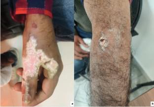
Results: A total of four isolates (clinical, n = 3; soil, n = 1) identified as Cladosporium halotolerans were included in the study. The clinical sites were skin, maxillary tissue and nail. All patients were apparently immunocompetent, and history of trauma was recorded in one patient. All patients improved on antifungal therapy. The cultures revealed growth of black mycelial fungus and microscopic examination demonstrated dematiaceous septate hyphae with erect conidiophores and conidia in branched acropetal chains. Based on molecular methods, all the four isolates were identified as C. halotolerans. SEM revealed no variation in length and width of the conidia, conidiophores, ramoconidium and hyphae among the isolates. All molecular targets, such as ITS region, LSU (partially sequenced), ACT and TUB were able to differentiate the isolates. Minimum inhibitory concentrations for antifungals were: triazoles (0.12-2 μg/ml), amphotericin B (4 μg/ml) and echinocandins (2-8 μg/ml).
Conclusion: We report role of the rarely isolated dematiaceous fungus, C. halotolerans, in causing human infections. The study emphasizes the role of molecular methods in precisely identifying these species. Triazoles are more active against these black fungi compared to polyenes or echinocandins.
期刊介绍:
Mycopathologia is an official journal of the International Union of Microbiological Societies (IUMS). Mycopathologia was founded in 1938 with the mission to ‘diffuse the understanding of fungal diseases in man and animals among mycologists’. Many of the milestones discoveries in the field of medical mycology have been communicated through the pages of this journal. Mycopathologia covers a diverse, interdisciplinary range of topics that is unique in breadth and depth. The journal publishes peer-reviewed, original articles highlighting important developments concerning medically important fungi and fungal diseases. The journal highlights important developments in fungal systematics and taxonomy, laboratory diagnosis of fungal infections, antifungal drugs, clinical presentation and treatment, and epidemiology of fungal diseases globally. Timely opinion articles, mini-reviews, and other communications are usually invited at the discretion of the editorial board. Unique case reports highlighting unprecedented progress in the diagnosis and treatment of fungal infections, are published in every issue of the journal. MycopathologiaIMAGE is another regular feature for a brief clinical report of potential interest to a mixed audience of physicians and laboratory scientists. MycopathologiaGENOME is designed for the rapid publication of new genomes of human and animal pathogenic fungi using a checklist-based, standardized format.
 求助内容:
求助内容: 应助结果提醒方式:


