Mamiko Takemoto, Yuta Kitamura, Masato Kakisu, Daisuke Shimizu, Takayuki Baba
{"title":"Retinal Pigment Epithelial Tears after Ex-PRESS Filtration Surgery in a Glaucoma Patient with a History of Ischemic Optic Neuropathy.","authors":"Mamiko Takemoto, Yuta Kitamura, Masato Kakisu, Daisuke Shimizu, Takayuki Baba","doi":"10.1155/2023/6645156","DOIUrl":null,"url":null,"abstract":"<p><strong>Background: </strong>To describe a case of retinal pigment epithelial tears (RPE tears) and serous retinal detachment (SRD) after Ex-PRESS filtration surgery for primary open-angle glaucoma (POAG) combined with ischemic optic neuropathy. <i>Case Presentation</i>. This case report involved a 69-year-old woman who underwent Ex-PRESS filtration surgery for right POAG. She had a history of systemic arteriosclerotic disease and subacute progressive visual field loss due to suspected ischemic optic neuropathy in her right eye. The right preoperative visual acuity was 0.7, and intraocular pressure (IOP) was 19 mmHg with maximum glaucoma eye drops. RPE detachment was not observed in the fundus. On day 9 after surgery, the IOP was 6 mmHg, and mild choroidal detachment was observed. On day 13, although IOP remained almost unchanged at 7 mmHg, bullous SRD was observed in the inferior retina, including the macula, and RPE tears were observed along the superior arcade vessel. While subretinal fluid gradually decreased with increasing IOP, tractional retinal folds persisted along the superior arcade, accompanied by macular degeneration.</p><p><strong>Conclusion: </strong>We experienced a case of RPE tears after Ex-PRESS filtration surgery. In addition to choroidal detachment in the setting of hypotony, a pathologic condition causing structural fragility of the RPE layer may contribute to the development of RPE tears.</p>","PeriodicalId":9603,"journal":{"name":"Case Reports in Ophthalmological Medicine","volume":"2023 ","pages":"6645156"},"PeriodicalIF":0.4000,"publicationDate":"2023-10-25","publicationTypes":"Journal Article","fieldsOfStudy":null,"isOpenAccess":false,"openAccessPdf":"https://www.ncbi.nlm.nih.gov/pmc/articles/PMC10620019/pdf/","citationCount":"0","resultStr":null,"platform":"Semanticscholar","paperid":null,"PeriodicalName":"Case Reports in Ophthalmological Medicine","FirstCategoryId":"1085","ListUrlMain":"https://doi.org/10.1155/2023/6645156","RegionNum":0,"RegionCategory":null,"ArticlePicture":[],"TitleCN":null,"AbstractTextCN":null,"PMCID":null,"EPubDate":"2023/1/1 0:00:00","PubModel":"eCollection","JCR":"Q4","JCRName":"OPHTHALMOLOGY","Score":null,"Total":0}
引用次数: 0
Abstract
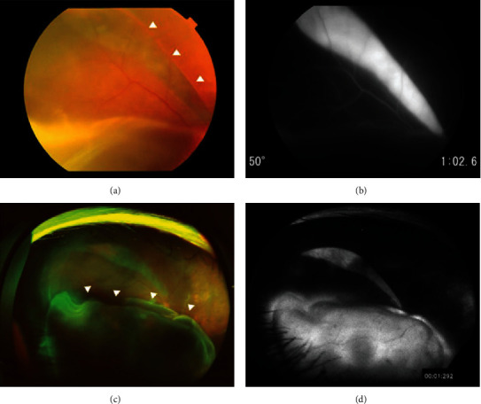
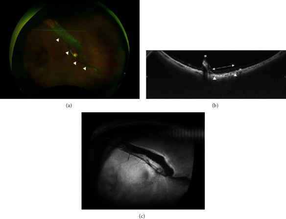
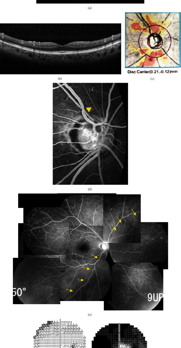
Background: To describe a case of retinal pigment epithelial tears (RPE tears) and serous retinal detachment (SRD) after Ex-PRESS filtration surgery for primary open-angle glaucoma (POAG) combined with ischemic optic neuropathy. Case Presentation. This case report involved a 69-year-old woman who underwent Ex-PRESS filtration surgery for right POAG. She had a history of systemic arteriosclerotic disease and subacute progressive visual field loss due to suspected ischemic optic neuropathy in her right eye. The right preoperative visual acuity was 0.7, and intraocular pressure (IOP) was 19 mmHg with maximum glaucoma eye drops. RPE detachment was not observed in the fundus. On day 9 after surgery, the IOP was 6 mmHg, and mild choroidal detachment was observed. On day 13, although IOP remained almost unchanged at 7 mmHg, bullous SRD was observed in the inferior retina, including the macula, and RPE tears were observed along the superior arcade vessel. While subretinal fluid gradually decreased with increasing IOP, tractional retinal folds persisted along the superior arcade, accompanied by macular degeneration.
Conclusion: We experienced a case of RPE tears after Ex-PRESS filtration surgery. In addition to choroidal detachment in the setting of hypotony, a pathologic condition causing structural fragility of the RPE layer may contribute to the development of RPE tears.

 求助内容:
求助内容: 应助结果提醒方式:


