Min Pan, Miao-Miao Zhang, Shu-Qin Xu, Yi Lyu, Xiao-Peng Yan
{"title":"Magnetic anchor technique assisted endoscopic submucosal dissection for early esophageal cancer.","authors":"Min Pan, Miao-Miao Zhang, Shu-Qin Xu, Yi Lyu, Xiao-Peng Yan","doi":"10.4253/wjge.v15.i10.584","DOIUrl":null,"url":null,"abstract":"<p><strong>Background: </strong>Esophageal cancer has high incidence globally and is often diagnosed at an advanced stage. With the widespread application of endoscopic technologies, the need for early detection and diagnosis of esophageal cancer has gradually been realized. Endoscopic submucosal dissection (ESD) has become the standard of care for managing early tumors of the esophagus, stomach, and colon. However, due to the steep learning curve, difficult operation, and technically demanding nature of the procedure, ESD has currently been committed to the development of various assistive technologies.</p><p><strong>Aim: </strong>To explore the feasibility and applicability of magnetic anchor technique (MAT)-assisted ESD for early esophageal cancer.</p><p><strong>Methods: </strong>Isolated pig esophagi were used as the experimental model, and the magnetic anchor device was designed by us. The esophagi used were divided into two groups, namely the operational and control groups, and 10 endoscopists completed the procedure. The two groups were evaluated for the following aspects: The total operative time, perforation rate, rate of whole mucosal resection, diameter of the peering mucosa, and scores of endoscopists' feelings with the procedure, including the convenience, mucosal surface exposure degree, and tissue tension. In addition, in the operational group, the soft tissue clip and the target magnet (TM) were connected by a thin wire through a small hole at the tail end of the TM. Under gastroscopic guidance, the soft tissue clip was clamped to the edge of the lesioned mucosa, which was marked in advance. By changing the position of the anchor magnet (AM) outside the esophagus, the pulling force and pulling direction of the TM could be changed, thus exposing the mucosal peeling surface and assisting the ESD.</p><p><strong>Results: </strong>Herein, each of the two groups comprised 10 isolated esophageal putative mucosal lesions. The diameter of the peering mucosa did not significantly differ between the two groups (2.13 ± 0.06 <i>vs</i> 2.15 ± 0.06, <i>P</i> = 0.882). The total operative time was shorter in the operational group than in the control group (17.04 ± 0.22 min <i>vs</i> 21.94 ± 0.23 min, <i>P</i> < 0.001). During the entire experiment, the TM remained firmly connected with the soft tissue clip and did not affect the opening, closing, and release of the soft tissue clip. The interaction between the TM and AM could provide sufficient tissue tension and completely expose the mucosa, which greatly assists the surgeon with the operation. There was no avulsion of the mucosa, and mucosal lesions were intact when peeled. Therefore, the scores of endoscopists' feelings were higher in the operational group than in the control group in terms of the convenience (9.22 ± 0.19 <i>vs</i> 8.34 ± 0.15, <i>P</i> = 0.002), mucosal surface exposure degree (9.11 ± 0.15 <i>vs</i> 8.25 ± 0.12, <i>P</i> < 0.001), and tissue tension (9.35 ± 0.13 <i>vs</i> 8.02 ± 0.17, <i>P</i> < 0.001). The two groups did not significantly differ in the perforation rate and rate of whole mucosal resection.</p><p><strong>Conclusion: </strong>We found MAT-assisted ESD safe and feasible for early esophageal cancer. It could greatly improve the endoscopic operation experience and showed good clinical application prospects.</p>","PeriodicalId":23953,"journal":{"name":"World Journal of Gastrointestinal Endoscopy","volume":"15 10","pages":"584-592"},"PeriodicalIF":1.4000,"publicationDate":"2023-10-16","publicationTypes":"Journal Article","fieldsOfStudy":null,"isOpenAccess":false,"openAccessPdf":"https://www.ncbi.nlm.nih.gov/pmc/articles/PMC10600693/pdf/","citationCount":"0","resultStr":null,"platform":"Semanticscholar","paperid":null,"PeriodicalName":"World Journal of Gastrointestinal Endoscopy","FirstCategoryId":"3","ListUrlMain":"https://doi.org/10.4253/wjge.v15.i10.584","RegionNum":0,"RegionCategory":null,"ArticlePicture":[],"TitleCN":null,"AbstractTextCN":null,"PMCID":null,"EPubDate":"","PubModel":"","JCR":"Q4","JCRName":"GASTROENTEROLOGY & HEPATOLOGY","Score":null,"Total":0}
引用次数: 0
Abstract
Background: Esophageal cancer has high incidence globally and is often diagnosed at an advanced stage. With the widespread application of endoscopic technologies, the need for early detection and diagnosis of esophageal cancer has gradually been realized. Endoscopic submucosal dissection (ESD) has become the standard of care for managing early tumors of the esophagus, stomach, and colon. However, due to the steep learning curve, difficult operation, and technically demanding nature of the procedure, ESD has currently been committed to the development of various assistive technologies.
Aim: To explore the feasibility and applicability of magnetic anchor technique (MAT)-assisted ESD for early esophageal cancer.
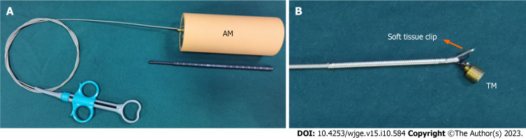
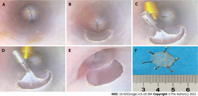
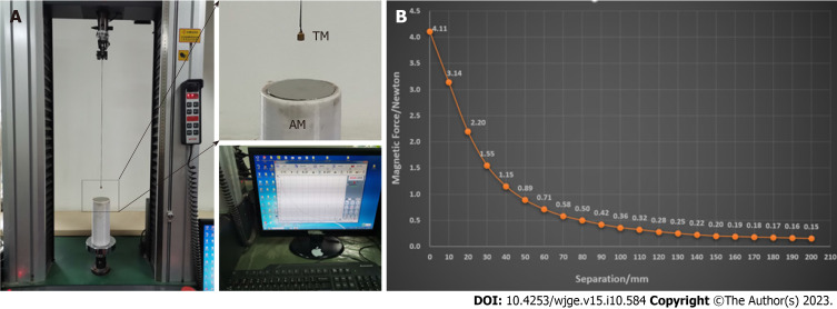
Methods: Isolated pig esophagi were used as the experimental model, and the magnetic anchor device was designed by us. The esophagi used were divided into two groups, namely the operational and control groups, and 10 endoscopists completed the procedure. The two groups were evaluated for the following aspects: The total operative time, perforation rate, rate of whole mucosal resection, diameter of the peering mucosa, and scores of endoscopists' feelings with the procedure, including the convenience, mucosal surface exposure degree, and tissue tension. In addition, in the operational group, the soft tissue clip and the target magnet (TM) were connected by a thin wire through a small hole at the tail end of the TM. Under gastroscopic guidance, the soft tissue clip was clamped to the edge of the lesioned mucosa, which was marked in advance. By changing the position of the anchor magnet (AM) outside the esophagus, the pulling force and pulling direction of the TM could be changed, thus exposing the mucosal peeling surface and assisting the ESD.
Results: Herein, each of the two groups comprised 10 isolated esophageal putative mucosal lesions. The diameter of the peering mucosa did not significantly differ between the two groups (2.13 ± 0.06 vs 2.15 ± 0.06, P = 0.882). The total operative time was shorter in the operational group than in the control group (17.04 ± 0.22 min vs 21.94 ± 0.23 min, P < 0.001). During the entire experiment, the TM remained firmly connected with the soft tissue clip and did not affect the opening, closing, and release of the soft tissue clip. The interaction between the TM and AM could provide sufficient tissue tension and completely expose the mucosa, which greatly assists the surgeon with the operation. There was no avulsion of the mucosa, and mucosal lesions were intact when peeled. Therefore, the scores of endoscopists' feelings were higher in the operational group than in the control group in terms of the convenience (9.22 ± 0.19 vs 8.34 ± 0.15, P = 0.002), mucosal surface exposure degree (9.11 ± 0.15 vs 8.25 ± 0.12, P < 0.001), and tissue tension (9.35 ± 0.13 vs 8.02 ± 0.17, P < 0.001). The two groups did not significantly differ in the perforation rate and rate of whole mucosal resection.
Conclusion: We found MAT-assisted ESD safe and feasible for early esophageal cancer. It could greatly improve the endoscopic operation experience and showed good clinical application prospects.
背景:癌症在全球范围内发病率很高,通常诊断为晚期。随着内镜技术的广泛应用,对癌症食管癌早期检测和诊断的需求逐渐实现。内镜下黏膜下剥离术(ESD)已成为治疗食道、胃和结肠早期肿瘤的标准护理方法。然而,由于该程序的学习曲线陡峭、操作困难和技术要求高,ESD目前致力于开发各种辅助技术。目的:探讨磁锚技术(MAT)辅助ESD治疗早期食管癌症的可行性和适用性。方法:以离体猪食管为实验模型,自行设计磁锚装置,将所用食管分为手术组和对照组,由10名内镜医生完成手术。对两组患者进行了以下方面的评估:总手术时间、穿孔率、全粘膜切除率、对等粘膜的直径以及内镜医生对手术的感受评分,包括方便性、粘膜表面暴露程度和组织张力。此外,在手术组中,软组织夹和靶磁体(TM)通过TM末端的小孔用细线连接。在胃镜引导下,将软组织夹夹在病变粘膜的边缘,预先做好标记。通过改变锚磁体(AM)在食管外的位置,可以改变TM的拉力和牵拉方向,从而暴露粘膜剥离表面,有助于ESD。两组对等粘膜的直径没有显著差异(2.13±0.06 vs 2.15±0.06,P=0.082)。手术组的总手术时间比对照组短(17.04±0.22分钟vs 21.94±0.23分钟,P<0.001)。在整个实验过程中,TM与软组织夹保持牢固连接,以及释放软组织夹。TM和AM之间的相互作用可以提供足够的组织张力并完全暴露粘膜,这大大有助于外科医生进行手术。粘膜没有撕脱,剥离时粘膜损伤完好无损。因此,在方便性(9.22±0.19 vs 8.34±0.15,P=0.002)、粘膜表面暴露程度(9.11±0.15 vs 8.25±0.12,P<0.001)、,组织张力(9.35±0.13 vs 8.02±0.17,P<0.001)。两组在穿孔率和全粘膜切除率方面没有显著差异。结论:MAT辅助ESD治疗早期食管癌症安全可行。它可以极大地提高内镜手术经验,具有良好的临床应用前景。
 求助内容:
求助内容: 应助结果提醒方式:


