{"title":"A Light and Ultrastructural Analysis in Odontogenic Cysts and Oral Squamous Cell Carcinoma.","authors":"Tanvi Handa, Shally Gupta, Simranjit Singh, Anubha Gulati","doi":"10.4103/jmau.jmau_64_23","DOIUrl":null,"url":null,"abstract":"<p><strong>Background: </strong>The aim of the study was to evaluate the desmosomal and hemidesmosomal attachments in common odontogenic cysts (radicular cyst [RC], odontogenic keratocyst [OKC], and dentigerous cyst [DC]), oral fibroma, and oral squamous cell carcinoma (OSCC). The disruption pattern in OKC was also compared with that of OSCC.</p><p><strong>Materials and methods: </strong>The 125 subjects included in the study were equally divided into five study groups - oral fibroma (Group I - control group), RC (Group II - test group), DC (Group III - test group), OKC (Group IV - test group), and OSCC (Group V - positive control). The clinical diagnosis was confirmed by histopathology, and the disruption was accessed through scanning electron microscopic images.</p><p><strong>Results: </strong>Oral fibroma displayed maximum intact desmosomes and hemidesmosomes and minimal disruption while in OSCC minimum intact desmosomes and hemidesmosomes were evident. Amongst the odontogenic cysts studied, OKC displayed maximum disrupted desmosomes and hemidesmsomes. Further, when OKC and OSCC were compared the completely intact desmosomes and hemidesmosomes were more in OKC than OSCC. The <i>P</i> value was set at <0.05.</p><p><strong>Conclusion: </strong>The study revealed that even though the defect in oral fibroma lies in the connective tissue, trauma or irritation as the etiology likely leads to minimal disruption in these intercellular junctions. These cell junctions were less evident in the case of DC owing to compression of the epithelial lining. The disruption of junctions in radicular cysts was more than those seen in oral fibroma. Compared to the other two cysts, OKCs displayed a much higher proportion of disruption in these cell junctions reflective of their more aggressive clinical behavior. OSCC displayed maximum disruption of cell junctions, which indicated that these disruptions play a role in both carcinogenesis and tumor invasion.</p>","PeriodicalId":16340,"journal":{"name":"Journal of Microscopy and Ultrastructure","volume":"1 1","pages":"142-150"},"PeriodicalIF":0.0000,"publicationDate":"2023-09-08","publicationTypes":"Journal Article","fieldsOfStudy":null,"isOpenAccess":false,"openAccessPdf":"https://www.ncbi.nlm.nih.gov/pmc/articles/PMC12499933/pdf/","citationCount":"0","resultStr":null,"platform":"Semanticscholar","paperid":null,"PeriodicalName":"Journal of Microscopy and Ultrastructure","FirstCategoryId":"1085","ListUrlMain":"https://doi.org/10.4103/jmau.jmau_64_23","RegionNum":0,"RegionCategory":null,"ArticlePicture":[],"TitleCN":null,"AbstractTextCN":null,"PMCID":null,"EPubDate":"2025/7/1 0:00:00","PubModel":"eCollection","JCR":"Q3","JCRName":"Medicine","Score":null,"Total":0}
引用次数: 0
Abstract
Background: The aim of the study was to evaluate the desmosomal and hemidesmosomal attachments in common odontogenic cysts (radicular cyst [RC], odontogenic keratocyst [OKC], and dentigerous cyst [DC]), oral fibroma, and oral squamous cell carcinoma (OSCC). The disruption pattern in OKC was also compared with that of OSCC.
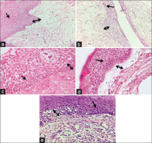
Materials and methods: The 125 subjects included in the study were equally divided into five study groups - oral fibroma (Group I - control group), RC (Group II - test group), DC (Group III - test group), OKC (Group IV - test group), and OSCC (Group V - positive control). The clinical diagnosis was confirmed by histopathology, and the disruption was accessed through scanning electron microscopic images.
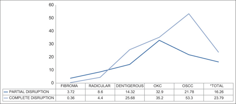
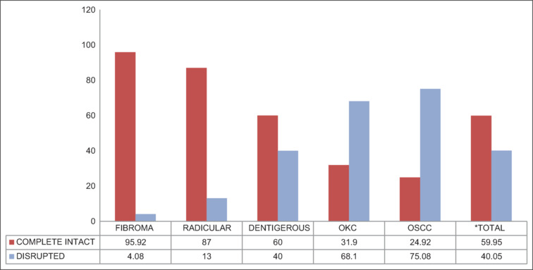
Results: Oral fibroma displayed maximum intact desmosomes and hemidesmosomes and minimal disruption while in OSCC minimum intact desmosomes and hemidesmosomes were evident. Amongst the odontogenic cysts studied, OKC displayed maximum disrupted desmosomes and hemidesmsomes. Further, when OKC and OSCC were compared the completely intact desmosomes and hemidesmosomes were more in OKC than OSCC. The P value was set at <0.05.
Conclusion: The study revealed that even though the defect in oral fibroma lies in the connective tissue, trauma or irritation as the etiology likely leads to minimal disruption in these intercellular junctions. These cell junctions were less evident in the case of DC owing to compression of the epithelial lining. The disruption of junctions in radicular cysts was more than those seen in oral fibroma. Compared to the other two cysts, OKCs displayed a much higher proportion of disruption in these cell junctions reflective of their more aggressive clinical behavior. OSCC displayed maximum disruption of cell junctions, which indicated that these disruptions play a role in both carcinogenesis and tumor invasion.

 求助内容:
求助内容: 应助结果提醒方式:


