{"title":"Alpha Thalassemia/Intellectual Disability Syndrome X-Linked Expression Varies Significantly between Androgen Receptor-Positive and Androgen Receptor-Negative Prostatic Adenocarcinoma: Relation to Epidermal Growth Factor Receptor Expression and Clinicopathological Factors.","authors":"Marwa A Abd El-Azeem, Dina A Radi","doi":"10.4103/jmau.jmau_119_22","DOIUrl":null,"url":null,"abstract":"<p><strong>Background: </strong>Prostate cancer (PC) is the most common cancer and a leading cause of cancer-related mortality in men worldwide. It has become clear that signaling pathways that are implicated in prostatic carcinoma (PCa) initiation and propagation evolved through interactions of several factors. Alpha Thalassemia/Intellectual Disability syndrome X-linked (ATRX) is a chromatin remodeling protein that has an essential role in telomere stability. The androgen receptor (AR) and growth factors especially the epidermal growth factor receptor (EGFR) seem not to function independently in PCa proliferation. This work aimed to study the expression of ATRX in primary prostatic adenocarcinoma in relation to AR and EGFR expression and the significance of biomarkers expression to the known clinicopathological factors.</p><p><strong>Materials and methods: </strong>Eighty-two primary prostatic adenocarcinoma paraffin blocks were stained immunohistochemically with AR, ATRX, and EGFR polyclonal antibodies. PCa was divided into AR<sup>+/hi</sup> and AR<sup>-/lo</sup> depending on the percentage and intensity of stained cells regardless of AR heterogeneity. ATRX immunostaining was categorized into ATRX preserved expression or ATRX loss. EGFR expression was grouped into low and high expression according to the staining percentage and intensity.</p><p><strong>Results: </strong>AR<sup>+/hi</sup> and preserved ATRX expression significantly were linked to low pT stage, low-grade group, and absence of lymph node invasion. While significant EGFR high expression was related to the high-grade group and the presence of lymph node invasion.</p><p><strong>Conclusion: </strong>ATRX preserved expression varies significantly between AR<sup>+/hi</sup> and AR<sup>-/lo</sup> PCa which is related to favorable clinicopathological factors. However, the loss of ATRX expression correlated significantly with AR<sup>-/low</sup>, high EGFR expression, and adverse clinicopathological factors.</p>","PeriodicalId":16340,"journal":{"name":"Journal of Microscopy and Ultrastructure","volume":"1 1","pages":"57-67"},"PeriodicalIF":0.0000,"publicationDate":"2023-03-22","publicationTypes":"Journal Article","fieldsOfStudy":null,"isOpenAccess":false,"openAccessPdf":"https://www.ncbi.nlm.nih.gov/pmc/articles/PMC12236420/pdf/","citationCount":"0","resultStr":null,"platform":"Semanticscholar","paperid":null,"PeriodicalName":"Journal of Microscopy and Ultrastructure","FirstCategoryId":"1085","ListUrlMain":"https://doi.org/10.4103/jmau.jmau_119_22","RegionNum":0,"RegionCategory":null,"ArticlePicture":[],"TitleCN":null,"AbstractTextCN":null,"PMCID":null,"EPubDate":"2025/4/1 0:00:00","PubModel":"eCollection","JCR":"Q3","JCRName":"Medicine","Score":null,"Total":0}
引用次数: 0
Abstract
Background: Prostate cancer (PC) is the most common cancer and a leading cause of cancer-related mortality in men worldwide. It has become clear that signaling pathways that are implicated in prostatic carcinoma (PCa) initiation and propagation evolved through interactions of several factors. Alpha Thalassemia/Intellectual Disability syndrome X-linked (ATRX) is a chromatin remodeling protein that has an essential role in telomere stability. The androgen receptor (AR) and growth factors especially the epidermal growth factor receptor (EGFR) seem not to function independently in PCa proliferation. This work aimed to study the expression of ATRX in primary prostatic adenocarcinoma in relation to AR and EGFR expression and the significance of biomarkers expression to the known clinicopathological factors.
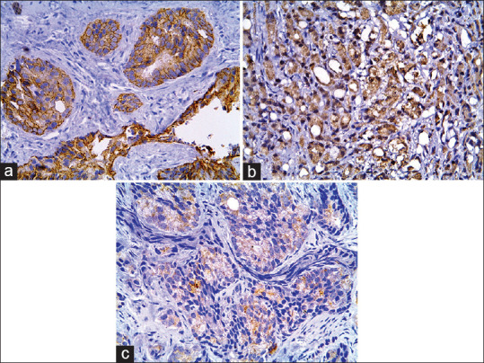
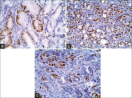
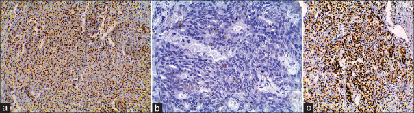
Materials and methods: Eighty-two primary prostatic adenocarcinoma paraffin blocks were stained immunohistochemically with AR, ATRX, and EGFR polyclonal antibodies. PCa was divided into AR+/hi and AR-/lo depending on the percentage and intensity of stained cells regardless of AR heterogeneity. ATRX immunostaining was categorized into ATRX preserved expression or ATRX loss. EGFR expression was grouped into low and high expression according to the staining percentage and intensity.
Results: AR+/hi and preserved ATRX expression significantly were linked to low pT stage, low-grade group, and absence of lymph node invasion. While significant EGFR high expression was related to the high-grade group and the presence of lymph node invasion.
Conclusion: ATRX preserved expression varies significantly between AR+/hi and AR-/lo PCa which is related to favorable clinicopathological factors. However, the loss of ATRX expression correlated significantly with AR-/low, high EGFR expression, and adverse clinicopathological factors.

 求助内容:
求助内容: 应助结果提醒方式:


