{"title":"Bioinspired Hydrogel Electrospun Fibers for Spinal Cord Regeneration","authors":"Chunmao Chen, Jincheng Tang, Yong Gu, Lili Liu, Xingzhi Liu, Lianfu Deng, Cláudia Martins, Bruno Sarmento, Wenguo Cui, Liang Chen","doi":"10.1002/adfm.201806899","DOIUrl":null,"url":null,"abstract":"<p>Fully simulating the components and microstructures of soft tissue is a challenge for its functional regeneration. A new aligned hydrogel microfiber scaffold for spinal cord regeneration is constructed with photocrosslinked gelatin methacryloyl (GelMA) and electrospinning technology. The directional porous hydrogel fibrous scaffold consistent with nerve axons is vital to guide cell migration and axon extension. The GelMA hydrogel electrospun fibers soak up water more than six times their weight, with a lower Young's modulus, providing a favorable survival and metabolic environment for neuronal cells. GelMA fibers further demonstrate higher antinestin, anti-Tuj-1, antisynaptophysin, and anti-CD31 gene expression in neural stem cells, neuronal cells, synapses, and vascular endothelial cells, respectively. In contrast, anti-GFAP and anti-CS56 labeled astrocytes and glial scars of GelMA fibers are shown to be present in a lesser extent compared with gelatin fibers. The soft bionic scaffold constructed with electrospun GelMA hydrogel fibers not only facilitates the migration of neural stem cells and induces their differentiation into neuronal cells, but also inhibits the glial scar formation and promotes angiogenesis. Moreover, the scaffold with a high degree of elasticity can resist deformation without the protection of a bony spinal canal. The bioinspired aligned hydrogel microfiber proves to be efficient and versatile in triggering functional regeneration of the spinal cord.</p>","PeriodicalId":112,"journal":{"name":"Advanced Functional Materials","volume":"29 4","pages":""},"PeriodicalIF":18.5000,"publicationDate":"2018-12-02","publicationTypes":"Journal Article","fieldsOfStudy":null,"isOpenAccess":false,"openAccessPdf":"https://sci-hub-pdf.com/10.1002/adfm.201806899","citationCount":"123","resultStr":null,"platform":"Semanticscholar","paperid":null,"PeriodicalName":"Advanced Functional Materials","FirstCategoryId":"88","ListUrlMain":"https://onlinelibrary.wiley.com/doi/10.1002/adfm.201806899","RegionNum":1,"RegionCategory":"材料科学","ArticlePicture":[],"TitleCN":null,"AbstractTextCN":null,"PMCID":null,"EPubDate":"","PubModel":"","JCR":"Q1","JCRName":"CHEMISTRY, MULTIDISCIPLINARY","Score":null,"Total":0}
引用次数: 123
Abstract
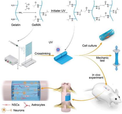
Fully simulating the components and microstructures of soft tissue is a challenge for its functional regeneration. A new aligned hydrogel microfiber scaffold for spinal cord regeneration is constructed with photocrosslinked gelatin methacryloyl (GelMA) and electrospinning technology. The directional porous hydrogel fibrous scaffold consistent with nerve axons is vital to guide cell migration and axon extension. The GelMA hydrogel electrospun fibers soak up water more than six times their weight, with a lower Young's modulus, providing a favorable survival and metabolic environment for neuronal cells. GelMA fibers further demonstrate higher antinestin, anti-Tuj-1, antisynaptophysin, and anti-CD31 gene expression in neural stem cells, neuronal cells, synapses, and vascular endothelial cells, respectively. In contrast, anti-GFAP and anti-CS56 labeled astrocytes and glial scars of GelMA fibers are shown to be present in a lesser extent compared with gelatin fibers. The soft bionic scaffold constructed with electrospun GelMA hydrogel fibers not only facilitates the migration of neural stem cells and induces their differentiation into neuronal cells, but also inhibits the glial scar formation and promotes angiogenesis. Moreover, the scaffold with a high degree of elasticity can resist deformation without the protection of a bony spinal canal. The bioinspired aligned hydrogel microfiber proves to be efficient and versatile in triggering functional regeneration of the spinal cord.
期刊介绍:
Firmly established as a top-tier materials science journal, Advanced Functional Materials reports breakthrough research in all aspects of materials science, including nanotechnology, chemistry, physics, and biology every week.
Advanced Functional Materials is known for its rapid and fair peer review, quality content, and high impact, making it the first choice of the international materials science community.
 求助内容:
求助内容: 应助结果提醒方式:


