{"title":"Quantifying the effects of long-range 13C-13C dipolar coupling on measured relaxation rates in RNA","authors":"Lukasz T. Olenginski, Theodore K. Dayie","doi":"10.1007/s10858-021-00368-8","DOIUrl":null,"url":null,"abstract":"<p>Selective stable isotope labeling has transformed structural and dynamics analysis of RNA by NMR spectroscopy. These methods can remove <sup>13</sup>C-<sup>13</sup>C dipolar couplings that complicate <sup>13</sup>C relaxation analyses. While these phenomena are well documented for sites with adjacent <sup>13</sup>C nuclei (e.g. ribose C1′), less is known about so-called isolated sites (e.g. adenosine C2). To investigate and quantify the effects of long-range (>?2??) <sup>13</sup>C-<sup>13</sup>C dipolar interactions on RNA dynamics, we simulated adenosine C2 relaxation rates in uniformly [U-<sup>13</sup>C/<sup>15</sup>N]-ATP or selectively [2-<sup>13</sup>C]-ATP labeled RNAs. Our simulations predict non-negligible <sup>13</sup>C-<sup>13</sup>C dipolar contributions from adenosine C4, C5, and C6 to C2 longitudinal (R<sub>1</sub>) relaxation rates in [U-<sup>13</sup>C/<sup>15</sup>N]-ATP labeled RNAs. Moreover, these contributions increase at higher magnetic fields and molecular weights to introduce discrepancies that exceed 50%. This will become increasingly important at GHz fields. Experimental R<sub>1</sub> measurements in the 61 nucleotide human hepatitis B virus encapsidation signal ε RNA labeled with [U-<sup>13</sup>C/<sup>15</sup>N]-ATP or [2-<sup>13</sup>C]-ATP corroborate these simulations. Thus, in the absence of selectively labeled samples, long-range <sup>13</sup>C-<sup>13</sup>C dipolar contributions must be explicitly taken into account when interpreting adenosine C2 R<sub>1</sub> rates in terms of motional models for large RNAs.</p>","PeriodicalId":613,"journal":{"name":"Journal of Biomolecular NMR","volume":"75 4-5","pages":"203 - 211"},"PeriodicalIF":1.3000,"publicationDate":"2021-04-29","publicationTypes":"Journal Article","fieldsOfStudy":null,"isOpenAccess":false,"openAccessPdf":"https://sci-hub-pdf.com/10.1007/s10858-021-00368-8","citationCount":"4","resultStr":null,"platform":"Semanticscholar","paperid":null,"PeriodicalName":"Journal of Biomolecular NMR","FirstCategoryId":"99","ListUrlMain":"https://link.springer.com/article/10.1007/s10858-021-00368-8","RegionNum":3,"RegionCategory":"生物学","ArticlePicture":[],"TitleCN":null,"AbstractTextCN":null,"PMCID":null,"EPubDate":"","PubModel":"","JCR":"Q3","JCRName":"BIOCHEMISTRY & MOLECULAR BIOLOGY","Score":null,"Total":0}
引用次数: 4
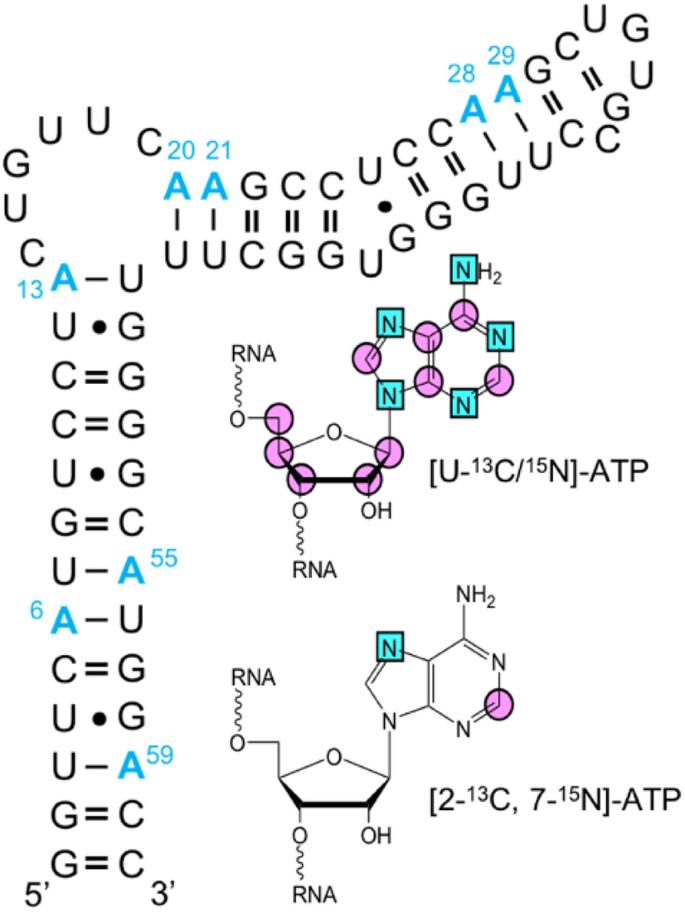
Abstract
Selective stable isotope labeling has transformed structural and dynamics analysis of RNA by NMR spectroscopy. These methods can remove 13C-13C dipolar couplings that complicate 13C relaxation analyses. While these phenomena are well documented for sites with adjacent 13C nuclei (e.g. ribose C1′), less is known about so-called isolated sites (e.g. adenosine C2). To investigate and quantify the effects of long-range (>?2??) 13C-13C dipolar interactions on RNA dynamics, we simulated adenosine C2 relaxation rates in uniformly [U-13C/15N]-ATP or selectively [2-13C]-ATP labeled RNAs. Our simulations predict non-negligible 13C-13C dipolar contributions from adenosine C4, C5, and C6 to C2 longitudinal (R1) relaxation rates in [U-13C/15N]-ATP labeled RNAs. Moreover, these contributions increase at higher magnetic fields and molecular weights to introduce discrepancies that exceed 50%. This will become increasingly important at GHz fields. Experimental R1 measurements in the 61 nucleotide human hepatitis B virus encapsidation signal ε RNA labeled with [U-13C/15N]-ATP or [2-13C]-ATP corroborate these simulations. Thus, in the absence of selectively labeled samples, long-range 13C-13C dipolar contributions must be explicitly taken into account when interpreting adenosine C2 R1 rates in terms of motional models for large RNAs.
期刊介绍:
The Journal of Biomolecular NMR provides a forum for publishing research on technical developments and innovative applications of nuclear magnetic resonance spectroscopy for the study of structure and dynamic properties of biopolymers in solution, liquid crystals, solids and mixed environments, e.g., attached to membranes. This may include:
Three-dimensional structure determination of biological macromolecules (polypeptides/proteins, DNA, RNA, oligosaccharides) by NMR.
New NMR techniques for studies of biological macromolecules.
Novel approaches to computer-aided automated analysis of multidimensional NMR spectra.
Computational methods for the structural interpretation of NMR data, including structure refinement.
Comparisons of structures determined by NMR with those obtained by other methods, e.g. by diffraction techniques with protein single crystals.
New techniques of sample preparation for NMR experiments (biosynthetic and chemical methods for isotope labeling, preparation of nutrients for biosynthetic isotope labeling, etc.). An NMR characterization of the products must be included.

 求助内容:
求助内容: 应助结果提醒方式:


