{"title":"Optical coherence tomography evaluation of deep dentin crack removal techniques","authors":"Daniel Hovander DDS , Grant Chyz DDS , Yasushi Shimada DDS, PhD , Junji Tagami DDS, PhD , Alireza Sadr DDS, PhD","doi":"10.1016/j.jfscie.2022.100012","DOIUrl":null,"url":null,"abstract":"<div><h3>Background</h3><p>This in vitro study used optical coherence tomography for noninvasive evaluation of the effectiveness of current clinical techniques to remove deep coronal dentin cracks.</p></div><div><h3>Methods</h3><p>Standard dentin cracks were induced on the pulpal floor of 40 decoronated, extracted, sound human posterior teeth using a diamond disk, resembling cracks extending from marginal ridges. The specimens were randomly assigned to 1 of 5 treatment groups for crack removal using airborne-particle abrasion or a bur (fissure, small round, medium round, or tapered fine diamond). Optical coherence tomographic scans were obtained before and after the crack removal. Three-dimensional image registration analyzed the amount of dentin removed and the dimensions of cracks initiated or propagated in each treatment group.</p></div><div><h3>Results</h3><p>Particle abrasion resulted in the smallest crack propagation in each dimension, which was significantly different from all bur groups (Mann-Whitney, <em>P</em> < .01). Removed dentin depth and width differed among burs (<em>P</em> < .05). All burs resulted in a degree of crack formation, and there was no difference in crack dimensions among bur groups (<em>P</em> > .05). The amount of removed dentin during crack treatment was the largest in the particle abrasion group, which was significantly different from those of the bur groups (Mann-Whitney, <em>P</em> < .005).</p></div><div><h3>Conclusions</h3><p>Dental burs used with the purpose of removing pulpal floor dentin cracks induced new small cracks and extended existing cracks, and the volume of removed dentin depended on the shape and size of the bur. Air particle abrasion induced the fewest new cracks despite removing larger dentin volume.</p></div>","PeriodicalId":73530,"journal":{"name":"JADA foundational science","volume":"1 ","pages":"Article 100012"},"PeriodicalIF":0.0000,"publicationDate":"2022-01-01","publicationTypes":"Journal Article","fieldsOfStudy":null,"isOpenAccess":false,"openAccessPdf":"https://www.sciencedirect.com/science/article/pii/S2772414X22000081/pdfft?md5=a524650487d6e993bd155e36dc176138&pid=1-s2.0-S2772414X22000081-main.pdf","citationCount":"0","resultStr":null,"platform":"Semanticscholar","paperid":null,"PeriodicalName":"JADA foundational science","FirstCategoryId":"1085","ListUrlMain":"https://www.sciencedirect.com/science/article/pii/S2772414X22000081","RegionNum":0,"RegionCategory":null,"ArticlePicture":[],"TitleCN":null,"AbstractTextCN":null,"PMCID":null,"EPubDate":"","PubModel":"","JCR":"","JCRName":"","Score":null,"Total":0}
引用次数: 0
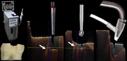
Abstract
Background
This in vitro study used optical coherence tomography for noninvasive evaluation of the effectiveness of current clinical techniques to remove deep coronal dentin cracks.
Methods
Standard dentin cracks were induced on the pulpal floor of 40 decoronated, extracted, sound human posterior teeth using a diamond disk, resembling cracks extending from marginal ridges. The specimens were randomly assigned to 1 of 5 treatment groups for crack removal using airborne-particle abrasion or a bur (fissure, small round, medium round, or tapered fine diamond). Optical coherence tomographic scans were obtained before and after the crack removal. Three-dimensional image registration analyzed the amount of dentin removed and the dimensions of cracks initiated or propagated in each treatment group.
Results
Particle abrasion resulted in the smallest crack propagation in each dimension, which was significantly different from all bur groups (Mann-Whitney, P < .01). Removed dentin depth and width differed among burs (P < .05). All burs resulted in a degree of crack formation, and there was no difference in crack dimensions among bur groups (P > .05). The amount of removed dentin during crack treatment was the largest in the particle abrasion group, which was significantly different from those of the bur groups (Mann-Whitney, P < .005).
Conclusions
Dental burs used with the purpose of removing pulpal floor dentin cracks induced new small cracks and extended existing cracks, and the volume of removed dentin depended on the shape and size of the bur. Air particle abrasion induced the fewest new cracks despite removing larger dentin volume.
本体外研究使用光学相干断层扫描对当前临床技术去除冠状牙本质深部裂缝的有效性进行无创评估。方法用金刚石盘在40颗人后牙的牙髓底诱导标准牙本质裂纹,形成从牙髓边缘脊向外延伸的裂纹。样本被随机分配到5个处理组中的1个,使用空气颗粒磨损或bur(裂纹,小圆,中圆或锥形细金刚石)去除裂纹。在裂纹去除前后分别进行了光学相干层析扫描。三维图像配准分析各组牙本质去除量和裂纹产生或扩展的尺寸。结果颗粒磨损导致各维度裂纹扩展最小,与所有磨损组差异显著(Mann-Whitney, P <. 01)。不同毛刺去除牙本质的深度和宽度不同(P <. 05)。所有毛刺都会形成一定程度的裂纹,不同毛刺组间裂纹尺寸没有差异(P >. 05)。裂纹处理过程中,颗粒磨损组的牙本质去除量最大,与bur组有显著差异(Mann-Whitney, P <.005)。结论牙髓底牙本质裂纹用牙髓刺去除时,牙本质裂纹产生新的小裂纹并扩展原有的裂纹,牙本质去除的体积取决于牙髓刺的形状和大小。空气颗粒磨损在去除较大牙本质体积的同时,产生的新裂纹最少。

 求助内容:
求助内容: 应助结果提醒方式:


