Victoria Armstrong, Cynthia Stretch, Liam Fitzgerald, Aquila Gopaul, Greg McKinnon, Jennifer Koziak, Karen Kopciuk, Nigel Brockton, Oliver F. Bathe
{"title":"Characterizing cancer-associated myosteatosis: anatomic distribution and cancer-specific variability of low radiodensity muscle","authors":"Victoria Armstrong, Cynthia Stretch, Liam Fitzgerald, Aquila Gopaul, Greg McKinnon, Jennifer Koziak, Karen Kopciuk, Nigel Brockton, Oliver F. Bathe","doi":"10.1002/rco2.46","DOIUrl":null,"url":null,"abstract":"<div>\n \n \n <section>\n \n <h3> Background</h3>\n \n <p>Low muscle radiodensity on computed tomography (CT) scan, indicative of myosteatosis, is commonly observed in cancer patients and can be associated with poor prognosis. Radiodensity is typically measured at the level of the third lumbar vertebra (L3). It is unknown whether features at L3 reflect a systemic state affecting peripheral muscle groups, whether images used at different levels can be used as a surrogate if L3 images are unavailable, and how radiodensity varies between cancer types.</p>\n </section>\n \n <section>\n \n <h3> Methods</h3>\n \n <p>Core and extremity muscle radiodensities were measured in whole body CT images from melanoma patients to evaluate the anatomical distribution of muscle radiodensity measurements. Core muscle radiodensity was measured in 891 patients with different cancer types to study malignancy-dependent patterns in muscle radiodensity.</p>\n </section>\n \n <section>\n \n <h3> Results</h3>\n \n <p>Low muscle radiodensity at L3 (<30 Hounsfield Unit) was associated with a corresponding lower muscle radiodensity in all muscle groups evaluated (<i>P</i> < 0.001). However, muscle radiodensities were lowest in the core muscle groups compared with muscles in the extremities. Muscle radiodensities at T12 closely correlated with measurements taken at L3 (<i>r</i> = 0.920, <i>P</i> < 0.001), but the correlation was weaker with mid-thigh measurements (<i>r</i> = 0.745, <i>P</i> < 0.001). The distribution of muscle radiodensities varied significantly with cancer type (<i>P</i> = 0.002).</p>\n </section>\n \n <section>\n \n <h3> Conclusions</h3>\n \n <p>The uniform distribution of low muscle radiodensity in cancer patients supports the hypothesis that the underlying mechanism for myosteatosis is systemic in nature. The most reliable measurements of muscle radiodensity are taken using images of core muscles. Variations in muscle radiodensity associated with cancer exist, suggesting that cancer-specific biological drivers are at play.</p>\n </section>\n </div>","PeriodicalId":73544,"journal":{"name":"JCSM rapid communications","volume":"4 2","pages":"197-206"},"PeriodicalIF":0.0000,"publicationDate":"2021-06-15","publicationTypes":"Journal Article","fieldsOfStudy":null,"isOpenAccess":false,"openAccessPdf":"https://sci-hub-pdf.com/10.1002/rco2.46","citationCount":"1","resultStr":null,"platform":"Semanticscholar","paperid":null,"PeriodicalName":"JCSM rapid communications","FirstCategoryId":"1085","ListUrlMain":"https://onlinelibrary.wiley.com/doi/10.1002/rco2.46","RegionNum":0,"RegionCategory":null,"ArticlePicture":[],"TitleCN":null,"AbstractTextCN":null,"PMCID":null,"EPubDate":"","PubModel":"","JCR":"","JCRName":"","Score":null,"Total":0}
引用次数: 1
Abstract
Background
Low muscle radiodensity on computed tomography (CT) scan, indicative of myosteatosis, is commonly observed in cancer patients and can be associated with poor prognosis. Radiodensity is typically measured at the level of the third lumbar vertebra (L3). It is unknown whether features at L3 reflect a systemic state affecting peripheral muscle groups, whether images used at different levels can be used as a surrogate if L3 images are unavailable, and how radiodensity varies between cancer types.
Methods
Core and extremity muscle radiodensities were measured in whole body CT images from melanoma patients to evaluate the anatomical distribution of muscle radiodensity measurements. Core muscle radiodensity was measured in 891 patients with different cancer types to study malignancy-dependent patterns in muscle radiodensity.
Results
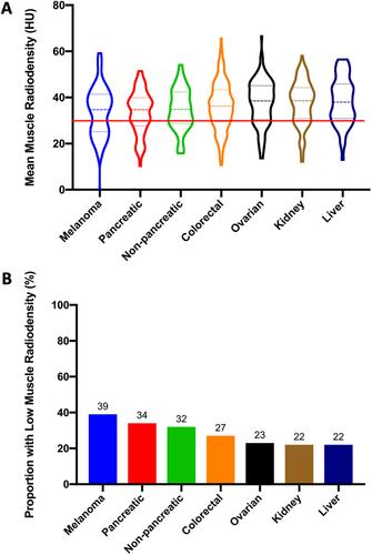
Low muscle radiodensity at L3 (<30 Hounsfield Unit) was associated with a corresponding lower muscle radiodensity in all muscle groups evaluated (P < 0.001). However, muscle radiodensities were lowest in the core muscle groups compared with muscles in the extremities. Muscle radiodensities at T12 closely correlated with measurements taken at L3 (r = 0.920, P < 0.001), but the correlation was weaker with mid-thigh measurements (r = 0.745, P < 0.001). The distribution of muscle radiodensities varied significantly with cancer type (P = 0.002).
Conclusions
The uniform distribution of low muscle radiodensity in cancer patients supports the hypothesis that the underlying mechanism for myosteatosis is systemic in nature. The most reliable measurements of muscle radiodensity are taken using images of core muscles. Variations in muscle radiodensity associated with cancer exist, suggesting that cancer-specific biological drivers are at play.

 求助内容:
求助内容: 应助结果提醒方式:


