Kishor R. Tardalkar , Leena R. Chaudhari , Mrunal N. Damle , Akshay A. Kawale , Nilesh C. Bhamare , Jeevitaa R. Kshersagar , Tanvee S. Kulkarni , Meghnad G. Joshi
{"title":"Extracellular-matrix Composite Bioink for 3D bioprinting and molding of small diameter vascular grafts","authors":"Kishor R. Tardalkar , Leena R. Chaudhari , Mrunal N. Damle , Akshay A. Kawale , Nilesh C. Bhamare , Jeevitaa R. Kshersagar , Tanvee S. Kulkarni , Meghnad G. Joshi","doi":"10.1016/j.bprint.2023.e00300","DOIUrl":null,"url":null,"abstract":"<div><p><span>Vascular grafts<span> are used in numerous vascular surgeries<span><span><span> around the world, although these procedures are constrained by vascular graft-related problems and size inconsistencies. This study developed and characterized vascular grafts using tissue engineering and </span>3D printing technology. To overcome vascular graft-related problems and size inconsistencies, this study developed a composite bio-ink using </span>ECM<span> of blood vessels, polyvinyl alcohol, and gelatin. Small-diameter vascular grafts were developed by Extrusion-based 3D printing and by molding techniques. The bioink was characterized in several aspects, followed by surface modification of a 3D vascular graft. The vascular grafts were evaluated for cytotoxicity and mechanical stability, which were found to be satisfactory. An </span></span></span></span><em>in vivo</em><span><span><span><span> biocompatibility and transplantation study showed cellular recruitment, </span>elastin<span> fibers, GAG as well as ECM (collagen) were retained. It was assessed by hematoxylin<span><span> and eosin (H&E), </span>alcian blue, and Masson's </span></span></span>trichrome staining. Recellularization and well-structured ECM were seen in </span>SEM<span> images. In immunohistochemistry<span>, positive vWF, α-SMA, and VEGF expression cells showed recruitment of endothelial and smooth muscle cells<span>. In conclusion, tissue specific bio-ink shows promise for further translational research and clinical application.</span></span></span></span></p></div>","PeriodicalId":37770,"journal":{"name":"Bioprinting","volume":null,"pages":null},"PeriodicalIF":0.0000,"publicationDate":"2023-10-01","publicationTypes":"Journal Article","fieldsOfStudy":null,"isOpenAccess":false,"openAccessPdf":"","citationCount":"0","resultStr":null,"platform":"Semanticscholar","paperid":null,"PeriodicalName":"Bioprinting","FirstCategoryId":"1085","ListUrlMain":"https://www.sciencedirect.com/science/article/pii/S240588662300043X","RegionNum":0,"RegionCategory":null,"ArticlePicture":[],"TitleCN":null,"AbstractTextCN":null,"PMCID":null,"EPubDate":"","PubModel":"","JCR":"Q1","JCRName":"Computer Science","Score":null,"Total":0}
引用次数: 0
Abstract
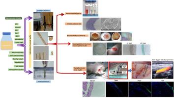
Vascular grafts are used in numerous vascular surgeries around the world, although these procedures are constrained by vascular graft-related problems and size inconsistencies. This study developed and characterized vascular grafts using tissue engineering and 3D printing technology. To overcome vascular graft-related problems and size inconsistencies, this study developed a composite bio-ink using ECM of blood vessels, polyvinyl alcohol, and gelatin. Small-diameter vascular grafts were developed by Extrusion-based 3D printing and by molding techniques. The bioink was characterized in several aspects, followed by surface modification of a 3D vascular graft. The vascular grafts were evaluated for cytotoxicity and mechanical stability, which were found to be satisfactory. An in vivo biocompatibility and transplantation study showed cellular recruitment, elastin fibers, GAG as well as ECM (collagen) were retained. It was assessed by hematoxylin and eosin (H&E), alcian blue, and Masson's trichrome staining. Recellularization and well-structured ECM were seen in SEM images. In immunohistochemistry, positive vWF, α-SMA, and VEGF expression cells showed recruitment of endothelial and smooth muscle cells. In conclusion, tissue specific bio-ink shows promise for further translational research and clinical application.
期刊介绍:
Bioprinting is a broad-spectrum, multidisciplinary journal that covers all aspects of 3D fabrication technology involving biological tissues, organs and cells for medical and biotechnology applications. Topics covered include nanomaterials, biomaterials, scaffolds, 3D printing technology, imaging and CAD/CAM software and hardware, post-printing bioreactor maturation, cell and biological factor patterning, biofabrication, tissue engineering and other applications of 3D bioprinting technology. Bioprinting publishes research reports describing novel results with high clinical significance in all areas of 3D bioprinting research. Bioprinting issues contain a wide variety of review and analysis articles covering topics relevant to 3D bioprinting ranging from basic biological, material and technical advances to pre-clinical and clinical applications of 3D bioprinting.

 求助内容:
求助内容: 应助结果提醒方式:


