{"title":"Supertemporal Resolution Imaging of Membrane Potential via Stroboscopic Microscopy","authors":"Luxin Peng, and , Peng Zou*, ","doi":"10.1021/cbmi.3c00054","DOIUrl":null,"url":null,"abstract":"<p >Membrane potential and its fluctuation are fundamental biophysical phenomena essential to cellular activities and functions. Compared to traditional electrode-based techniques, the optical recording via developed genetically encoded voltage indicators (GEVIs) offers a combination of noninvasiveness, high spatial resolution, and increased measurement throughput. However, its application is limited by the insufficient acquisition rate and time accuracy of the camera. Here we design and apply a stroboscopic illumination scheme to boost the temporal resolution of voltage imaging, while simultaneously eliminating the artifacts caused by nonsynchronized exposure in the rolling-shutter mode. We demonstrate that commonly used GEVIs are compatible with stroboscopic voltage imaging (SVI), and our SVI scheme offers a 5-fold faster acquisition frame rate than that of conventional continuous illumination. The GEVIs tested maintain high sensitivities in the SVI mode, supporting faithful reports of intracellular depolarization waveform and intercellular gap junction-mediated depolarization coupling in human embryonic kidney 293T (HEK 293T) cell populations. SVI allows resolving the action potential (AP) waveform with less distortion and mapping action potential initiation and propagation dynamics in cultured neurons in kilohertz, beyond the restriction from the camera in the field of view.</p>","PeriodicalId":53181,"journal":{"name":"Chemical & Biomedical Imaging","volume":null,"pages":null},"PeriodicalIF":0.0000,"publicationDate":"2023-07-20","publicationTypes":"Journal Article","fieldsOfStudy":null,"isOpenAccess":false,"openAccessPdf":"https://pubs.acs.org/doi/epdf/10.1021/cbmi.3c00054","citationCount":"0","resultStr":null,"platform":"Semanticscholar","paperid":null,"PeriodicalName":"Chemical & Biomedical Imaging","FirstCategoryId":"1085","ListUrlMain":"https://pubs.acs.org/doi/10.1021/cbmi.3c00054","RegionNum":0,"RegionCategory":null,"ArticlePicture":[],"TitleCN":null,"AbstractTextCN":null,"PMCID":null,"EPubDate":"","PubModel":"","JCR":"","JCRName":"","Score":null,"Total":0}
引用次数: 0
Abstract
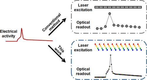
Membrane potential and its fluctuation are fundamental biophysical phenomena essential to cellular activities and functions. Compared to traditional electrode-based techniques, the optical recording via developed genetically encoded voltage indicators (GEVIs) offers a combination of noninvasiveness, high spatial resolution, and increased measurement throughput. However, its application is limited by the insufficient acquisition rate and time accuracy of the camera. Here we design and apply a stroboscopic illumination scheme to boost the temporal resolution of voltage imaging, while simultaneously eliminating the artifacts caused by nonsynchronized exposure in the rolling-shutter mode. We demonstrate that commonly used GEVIs are compatible with stroboscopic voltage imaging (SVI), and our SVI scheme offers a 5-fold faster acquisition frame rate than that of conventional continuous illumination. The GEVIs tested maintain high sensitivities in the SVI mode, supporting faithful reports of intracellular depolarization waveform and intercellular gap junction-mediated depolarization coupling in human embryonic kidney 293T (HEK 293T) cell populations. SVI allows resolving the action potential (AP) waveform with less distortion and mapping action potential initiation and propagation dynamics in cultured neurons in kilohertz, beyond the restriction from the camera in the field of view.
期刊介绍:
Chemical & Biomedical Imaging is a peer-reviewed open access journal devoted to the publication of cutting-edge research papers on all aspects of chemical and biomedical imaging. This interdisciplinary field sits at the intersection of chemistry physics biology materials engineering and medicine. The journal aims to bring together researchers from across these disciplines to address cutting-edge challenges of fundamental research and applications.Topics of particular interest include but are not limited to:Imaging of processes and reactionsImaging of nanoscale microscale and mesoscale materialsImaging of biological interactions and interfacesSingle-molecule and cellular imagingWhole-organ and whole-body imagingMolecular imaging probes and contrast agentsBioluminescence chemiluminescence and electrochemiluminescence imagingNanophotonics and imagingChemical tools for new imaging modalitiesChemical and imaging techniques in diagnosis and therapyImaging-guided drug deliveryAI and machine learning assisted imaging
 求助内容:
求助内容: 应助结果提醒方式:


