{"title":"A New Method for Optimizing the Size of Axial FOV in TOF-PEM to Improve Performance of the Scanner.","authors":"Delband Roshani, Saeed Setayeshi","doi":"10.31661/jbpe.v0i0.2009-1190","DOIUrl":null,"url":null,"abstract":"<p><strong>Background: </strong>Positron Emission Mammography (PEM) is a nuclear medicine imaging tool, playing a significant role in the diagnosis of patients with breast cancer. These days, many research has been done in order to improve the performance of this system.</p><p><strong>Objective: </strong>This study aims to propose a new method for optimizing the size of axial Field of View (FOV) in PEMs and improving the performance of the systems.</p><p><strong>Material and methods: </strong>In this analytical study, a conventional Inveon PET is simulated using GATE in order to validate the simulation. For this simulation, the mean relative difference is 2.91%, showing the precision and correction of simulation and consequently it is benchmarked. In the next step, for design of the new optimized detector, several validated simulations are performed in order to find the best geometry.</p><p><strong>Results: </strong>The best result is obtained with the axial FOV of 101.7 mm. It has 1.6×1.6×15 mm<sup>3</sup> lutetium yttrium orthosilicate (LYSO) crystals. The detector consists of 6 block rings with 30 detector blocks in each ring. In this paper, the performance of the scanner is improved and the geometry is optimized. Sensitivity and scatter fraction of the designed scanner are 4.65% and 21.2%, respectively, also noise equivalent count rate (NECR) is 105.442 kcps.</p><p><strong>Conclusion: </strong>The results showed 1 up to 3% improvement in the sensitivity of this new detector compared with different PEMs.</p>","PeriodicalId":38035,"journal":{"name":"Journal of Biomedical Physics and Engineering","volume":"13 5","pages":"471-476"},"PeriodicalIF":0.0000,"publicationDate":"2023-10-01","publicationTypes":"Journal Article","fieldsOfStudy":null,"isOpenAccess":false,"openAccessPdf":"https://ftp.ncbi.nlm.nih.gov/pub/pmc/oa_pdf/71/70/JBPE-13-471.PMC10589686.pdf","citationCount":"0","resultStr":null,"platform":"Semanticscholar","paperid":null,"PeriodicalName":"Journal of Biomedical Physics and Engineering","FirstCategoryId":"1085","ListUrlMain":"https://doi.org/10.31661/jbpe.v0i0.2009-1190","RegionNum":0,"RegionCategory":null,"ArticlePicture":[],"TitleCN":null,"AbstractTextCN":null,"PMCID":null,"EPubDate":"","PubModel":"","JCR":"Q3","JCRName":"Medicine","Score":null,"Total":0}
引用次数: 0
Abstract
Background: Positron Emission Mammography (PEM) is a nuclear medicine imaging tool, playing a significant role in the diagnosis of patients with breast cancer. These days, many research has been done in order to improve the performance of this system.
Objective: This study aims to propose a new method for optimizing the size of axial Field of View (FOV) in PEMs and improving the performance of the systems.
Material and methods: In this analytical study, a conventional Inveon PET is simulated using GATE in order to validate the simulation. For this simulation, the mean relative difference is 2.91%, showing the precision and correction of simulation and consequently it is benchmarked. In the next step, for design of the new optimized detector, several validated simulations are performed in order to find the best geometry.
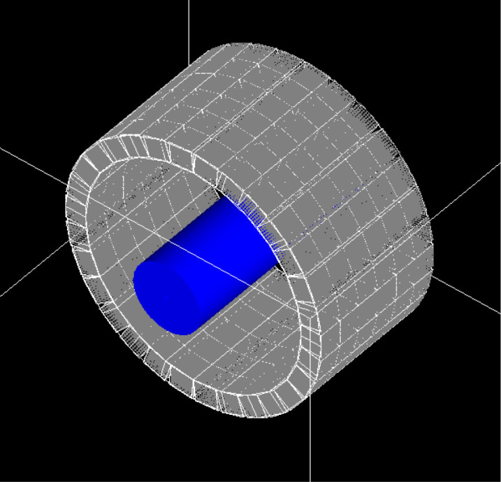
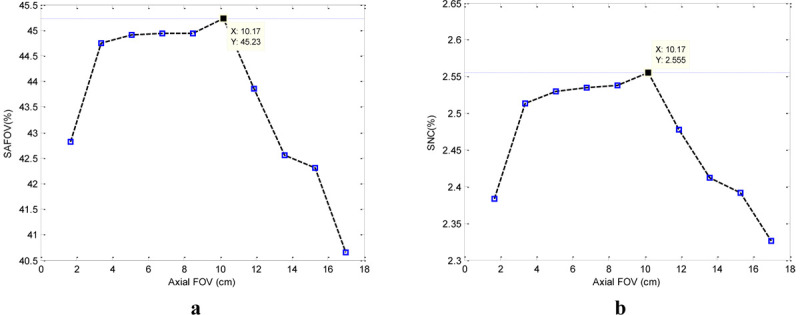
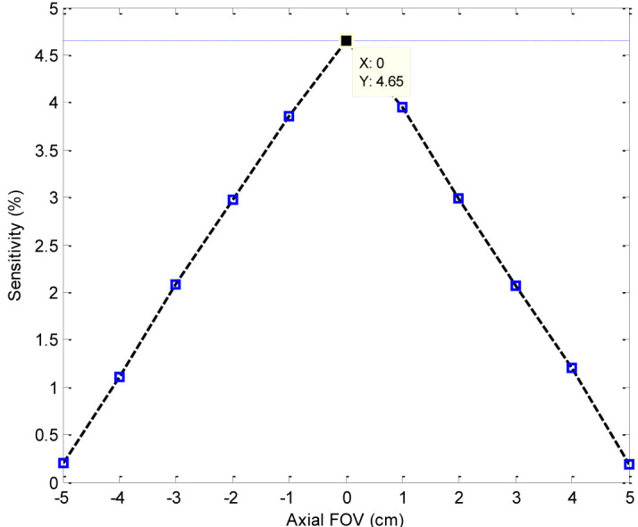
Results: The best result is obtained with the axial FOV of 101.7 mm. It has 1.6×1.6×15 mm3 lutetium yttrium orthosilicate (LYSO) crystals. The detector consists of 6 block rings with 30 detector blocks in each ring. In this paper, the performance of the scanner is improved and the geometry is optimized. Sensitivity and scatter fraction of the designed scanner are 4.65% and 21.2%, respectively, also noise equivalent count rate (NECR) is 105.442 kcps.
Conclusion: The results showed 1 up to 3% improvement in the sensitivity of this new detector compared with different PEMs.
期刊介绍:
The Journal of Biomedical Physics and Engineering (JBPE) is a bimonthly peer-reviewed English-language journal that publishes high-quality basic sciences and clinical research (experimental or theoretical) broadly concerned with the relationship of physics to medicine and engineering.

 求助内容:
求助内容: 应助结果提醒方式:


