Sangil Park, Boseong Kwon, Deok Hee Lee, Jae-Sung Ahn, Yunsun Song
{"title":"Ruptured Medullary Hemangioblastoma Mimicking a Craniocervical Junction Dural Arteriovenous Fistula with a Pseudoaneurysm.","authors":"Sangil Park, Boseong Kwon, Deok Hee Lee, Jae-Sung Ahn, Yunsun Song","doi":"10.5469/neuroint.2023.00451","DOIUrl":null,"url":null,"abstract":"<p><p>Hemangioblastomas (HBMs) are rare vascular tumors commonly located in the posterior fossa of adults. A mid-50s patient presented with sudden unconsciousness. Computed tomography scans revealed acute hemorrhages around the posterior fossa, predominantly in the subarachnoid space. Digital subtraction angiography (DSA) revealed an 8-mm round lesion filled with contrast agent, fed by the C1 segmental artery of the left vertebral artery (VA), showing early venous drainage to the spinal cord and brainstem. Emergent embolization was attempted under suspicion of a ruptured dural arteriovenous fistula, resulting in parent artery occlusion due to feeder selection failure. Follow-up DSA after a month depicted a persistent aneurysm via collaterals from both VAs. Consequently, the decision was made to proceed with surgical intervention, leading to the resection of the lesion, confirming its diagnosis as a HBM through histological examination. This case underscores the potential for misdiagnosis when HBMs with an intratumoral shunt mimic vascular shunt lesions.</p>","PeriodicalId":19140,"journal":{"name":"Neurointervention","volume":" ","pages":"209-213"},"PeriodicalIF":1.2000,"publicationDate":"2023-11-01","publicationTypes":"Journal Article","fieldsOfStudy":null,"isOpenAccess":false,"openAccessPdf":"https://www.ncbi.nlm.nih.gov/pmc/articles/PMC10626037/pdf/","citationCount":"0","resultStr":null,"platform":"Semanticscholar","paperid":null,"PeriodicalName":"Neurointervention","FirstCategoryId":"1085","ListUrlMain":"https://doi.org/10.5469/neuroint.2023.00451","RegionNum":0,"RegionCategory":null,"ArticlePicture":[],"TitleCN":null,"AbstractTextCN":null,"PMCID":null,"EPubDate":"2023/10/23 0:00:00","PubModel":"Epub","JCR":"Q4","JCRName":"CLINICAL NEUROLOGY","Score":null,"Total":0}
引用次数: 0
Abstract
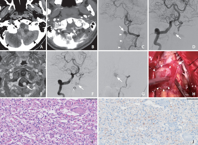
Hemangioblastomas (HBMs) are rare vascular tumors commonly located in the posterior fossa of adults. A mid-50s patient presented with sudden unconsciousness. Computed tomography scans revealed acute hemorrhages around the posterior fossa, predominantly in the subarachnoid space. Digital subtraction angiography (DSA) revealed an 8-mm round lesion filled with contrast agent, fed by the C1 segmental artery of the left vertebral artery (VA), showing early venous drainage to the spinal cord and brainstem. Emergent embolization was attempted under suspicion of a ruptured dural arteriovenous fistula, resulting in parent artery occlusion due to feeder selection failure. Follow-up DSA after a month depicted a persistent aneurysm via collaterals from both VAs. Consequently, the decision was made to proceed with surgical intervention, leading to the resection of the lesion, confirming its diagnosis as a HBM through histological examination. This case underscores the potential for misdiagnosis when HBMs with an intratumoral shunt mimic vascular shunt lesions.

 求助内容:
求助内容: 应助结果提醒方式:


