{"title":"Quantifying colors at micrometer scale by colorimetric microscopy (C-Microscopy) approach","authors":"Benedykt R. Jany","doi":"10.1016/j.micron.2023.103557","DOIUrl":null,"url":null,"abstract":"<div><p>The color is the primal property of the objects around us and is direct manifestation of light-matter interactions. The color information is used in many different fields of science, technology and industry to investigate material properties or for identification of concentrations of substances. Usually the color information is used as a global parameter in a macro scale. To quantitatively measure color information in micro scale one needs to use dedicated microscope spectrophotometers or specialized micro-reflectance setups. Here, the Colorimetric Microscopy (C-Microscopy) approach based on digital optical microscopy and a free software is presented. The C-Microscopy approach uses color calibrated image and colorimetric calculations to obtain physically meaningful quantities i.e., dominant wavelength and excitation purity maps at micro level scale. This allows for the discovery of the local color details of samples surfaces. Later, to fully characterize the optical properties, the hyperspectral reflectance data at micro scale (reflectance as a function of wavelength for a each point) are colorimetrically recovered. The C-Microscopy approach was successfully applied to various types of samples i.e., two metamorphic rocks unakite and lapis lazuli, which are mixtures of different minerals; and to the surface of gold 99.999 % pellet, which exhibits different types of surface features. The C-Microscopy approach could be used to quantify the local optical properties changes of various materials at microscale in an accessible way. The approach is freely available as a set of python jupyter notebooks.</p></div>","PeriodicalId":18501,"journal":{"name":"Micron","volume":null,"pages":null},"PeriodicalIF":2.5000,"publicationDate":"2023-10-14","publicationTypes":"Journal Article","fieldsOfStudy":null,"isOpenAccess":false,"openAccessPdf":"","citationCount":"0","resultStr":null,"platform":"Semanticscholar","paperid":null,"PeriodicalName":"Micron","FirstCategoryId":"5","ListUrlMain":"https://www.sciencedirect.com/science/article/pii/S0968432823001555","RegionNum":3,"RegionCategory":"工程技术","ArticlePicture":[],"TitleCN":null,"AbstractTextCN":null,"PMCID":null,"EPubDate":"","PubModel":"","JCR":"Q1","JCRName":"MICROSCOPY","Score":null,"Total":0}
引用次数: 0
Abstract
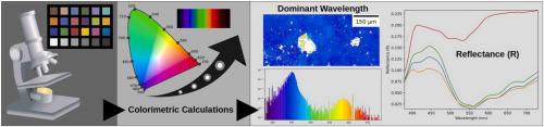
The color is the primal property of the objects around us and is direct manifestation of light-matter interactions. The color information is used in many different fields of science, technology and industry to investigate material properties or for identification of concentrations of substances. Usually the color information is used as a global parameter in a macro scale. To quantitatively measure color information in micro scale one needs to use dedicated microscope spectrophotometers or specialized micro-reflectance setups. Here, the Colorimetric Microscopy (C-Microscopy) approach based on digital optical microscopy and a free software is presented. The C-Microscopy approach uses color calibrated image and colorimetric calculations to obtain physically meaningful quantities i.e., dominant wavelength and excitation purity maps at micro level scale. This allows for the discovery of the local color details of samples surfaces. Later, to fully characterize the optical properties, the hyperspectral reflectance data at micro scale (reflectance as a function of wavelength for a each point) are colorimetrically recovered. The C-Microscopy approach was successfully applied to various types of samples i.e., two metamorphic rocks unakite and lapis lazuli, which are mixtures of different minerals; and to the surface of gold 99.999 % pellet, which exhibits different types of surface features. The C-Microscopy approach could be used to quantify the local optical properties changes of various materials at microscale in an accessible way. The approach is freely available as a set of python jupyter notebooks.
期刊介绍:
Micron is an interdisciplinary forum for all work that involves new applications of microscopy or where advanced microscopy plays a central role. The journal will publish on the design, methods, application, practice or theory of microscopy and microanalysis, including reports on optical, electron-beam, X-ray microtomography, and scanning-probe systems. It also aims at the regular publication of review papers, short communications, as well as thematic issues on contemporary developments in microscopy and microanalysis. The journal embraces original research in which microscopy has contributed significantly to knowledge in biology, life science, nanoscience and nanotechnology, materials science and engineering.
 求助内容:
求助内容: 应助结果提醒方式:


