Relationship between preoperative programmed death receptor 1, programmed death ligand 1 and clinical pathological parameters, early postoperative recurrence and metastasis in Patients with Esophageal Squamous Cell Carcinoma
引用次数: 0
Abstract
Objective To investigate the relationship between programmed death 1 (PD-1), programmed death receptor-1 ligand (PD-L1) and clinical pathological parameters, early postoperative recurrence and metastasis in patients with esophageal squamous cell carcinoma. Methods The retrospectively analyze of Paraffin tissue specimens and clinical pathology data in 58 Patients undergoing radical esophageal squamous cell carcinoma surgery from January 2015 to January 2017 in the 910 hospital of PLA Joint Service Support force were performed.Expression of PD-1 and PD-L1 in esophageal squamous cell carcinoma and normal esophageal mucosa were detected by SP immunohistochemical staining.The positive expression rates of PD-1 and PD-L1 in normal esophageal mucosa and esophageal squamous cell carcinoma were compared.the relationship between PD-1 and PD-L1 and gender, age, family history, depth of tumor invasion, degree of differentiation, lymph node metastasis, and TNM staging were analyzed.Follow-up was performed by outpatient consultation and telephone consultation.The recurrence and metastasis of early postoperative (≤1 year) was analyzed.The PD-1 and PD-L1 in esophageal squamous cell carcinoma were analyzed in patients with recurrent metastasis and non-relapsing and metastasis. Results The positive expression rate of PD-1 in esophageal squamous cell carcinoma was 37.93%(22/58), which was significantly higher than that in normal esophageal mucosa 15.52%(9/58). The difference was statistically significant (χ2=7.440, P=0.006). The positive expression rate of PD-L1 in esophageal squamous cell carcinoma was 43.10%(25/58), which was significantly higher than that of normal esophageal mucosa 18.97%(11/58). The difference was statistically significant (χ2=7.894, P=0.005). There was a difference in the positive expression rate of PD-L1 between different infiltration depth and TNM stage, P 0.05). The positive expression rate of PD-L1 in the recurrence group was 71.43% (10/14), and that in the non-recurrent group was 34.09% (15/44). The difference was statistically significant, (χ2=6.037, P<0.05). Conclusion The expression of PD-1 and PD-L1 in cancer tissues of patients with esophageal squamous cell carcinoma is highly expressed.PD-L1 is closely related to the occurrence and progression of esophageal squamous cell carcinoma, and it is also an important index affecting early recurrence and metastasis.Which can be selected as a new target for early diagnosis and treatment. Key words: Esophageal squamous cell carcinoma; Programmed death 1; Programmed death receptor-1 ligand; Clinicopathological features; Recurrence and metastasis; Correlation食管鳞状细胞癌术前程序性死亡受体1、程序性死亡配体1与临床病理参数及术后早期复发转移的关系
目的探讨程序性死亡1(PD-1)、程序性死亡受体1配体(PD-L1)与食管鳞状细胞癌临床病理参数、术后早期复发和转移的关系。方法回顾性分析2015年1月至2017年1月在中国人民解放军联勤保障部队910医院接受食管鳞状细胞癌根治术的58例患者的石蜡组织标本和临床病理资料。应用免疫组化SP法检测PD-1和PD-L1在食管鳞状细胞癌和正常食管黏膜中的表达。比较PD-1和PD-L1在正常食管黏膜和食管鳞状细胞癌中的阳性表达率,分析其与性别、年龄、家族史、肿瘤浸润深度、分化程度、淋巴结转移和TNM分期的关系。通过门诊咨询和电话咨询进行随访。分析术后早期(≤1年)的复发和转移情况。分析食管鳞状细胞癌复发转移和非复发转移患者的PD-1和PD-L1。结果PD-1在食管鳞状细胞癌中的阳性表达率为37.93%(22/58),明显高于正常食管粘膜中的15.52%(9/58),差异有统计学意义(χ2=7.440,P=0.006),差异有统计学意义(χ2=7.894,P=0.005),不同浸润深度和TNM分期的PD-L1阳性表达率有差异(P<0.05),复发组PD-L1阳性阳性表达率为71.43%(10/14),非复发组为34.09%(15/44)。结论PD-1和PD-L1在食管鳞状细胞癌癌症组织中表达较高。PD-L1与食管鳞状细胞癌的发生、发展密切相关,也是影响早期复发和转移的重要指标。可作为早期诊断和治疗的新靶点。关键词:食管鳞状细胞癌;程序性死亡1;程序性死亡受体-1配体;临床病理特征;复发和转移;相关性
本文章由计算机程序翻译,如有差异,请以英文原文为准。
求助全文
约1分钟内获得全文
求助全文
来源期刊
CiteScore
0.10
自引率
0.00%
发文量
16855
期刊介绍:
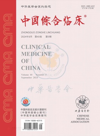
Clinical Medicine of China is an academic journal organized by the Chinese Medical Association (CMA), which mainly publishes original research papers, reviews and commentaries in the field.
Clinical Medicine of China is a source journal of Peking University (2000 and 2004 editions), a core journal of Chinese science and technology, an academic journal of RCCSE China Core (Extended Edition), and has been published in Chemical Abstracts of the United States (CA), Abstracts Journal of Russia (AJ), Chinese Core Journals (Selection) Database, Chinese Science and Technology Materials Directory, Wanfang Database, China Academic Journal Database, JST Japan Science and Technology Agency Database (Japanese) (2018) and other databases.

 求助内容:
求助内容: 应助结果提醒方式:


