{"title":"Retina regeneration: lessons from vertebrates.","authors":"Poonam Sharma, Rajesh Ramachandran","doi":"10.1093/oons/kvac012","DOIUrl":null,"url":null,"abstract":"<p><p>Unlike mammals, vertebrates such as fishes and frogs exhibit remarkable tissue regeneration including the central nervous system. Retina being part of the central nervous system has attracted the interest of several research groups to explore its regenerative ability in different vertebrate models including mice. Fishes and frogs completely restore the size, shape and tissue structure of an injured retina. Several studies have unraveled molecular mechanisms underlying retina regeneration. In teleosts, soon after injury, the Müller glial cells of the retina reprogram to form a proliferating population of Müller glia-derived progenitor cells capable of differentiating into various neural cell types and Müller glia. In amphibians, the transdifferentiation of retinal pigment epithelium and differentiation of ciliary marginal zone cells contribute to retina regeneration. In chicks and mice, supplementation with external growth factors or genetic modifications cause a partial regenerative response in the damaged retina. The initiation of retina regeneration is achieved through sequential orchestration of gene expression through controlled modulations in the genetic and epigenetic landscape of the progenitor cells. Several developmental biology pathways are turned on during the Müller glia reprogramming, retinal pigment epithelium transdifferentiation and ciliary marginal zone differentiation. Further, several tumorigenic pathways and gene expression events also contribute to the complete regeneration cascade of events. In this review, we address the various retinal injury paradigms and subsequent gene expression events governed in different vertebrate species. Further, we compared how vertebrates such as teleost fishes and amphibians can achieve excellent regenerative responses in the retina compared with their mammalian counterparts.</p>","PeriodicalId":74386,"journal":{"name":"Oxford open neuroscience","volume":" ","pages":"kvac012"},"PeriodicalIF":0.0000,"publicationDate":"2022-08-02","publicationTypes":"Journal Article","fieldsOfStudy":null,"isOpenAccess":false,"openAccessPdf":"https://www.ncbi.nlm.nih.gov/pmc/articles/PMC10913848/pdf/","citationCount":"0","resultStr":null,"platform":"Semanticscholar","paperid":null,"PeriodicalName":"Oxford open neuroscience","FirstCategoryId":"1085","ListUrlMain":"https://doi.org/10.1093/oons/kvac012","RegionNum":0,"RegionCategory":null,"ArticlePicture":[],"TitleCN":null,"AbstractTextCN":null,"PMCID":null,"EPubDate":"2022/1/1 0:00:00","PubModel":"eCollection","JCR":"","JCRName":"","Score":null,"Total":0}
引用次数: 0
Abstract
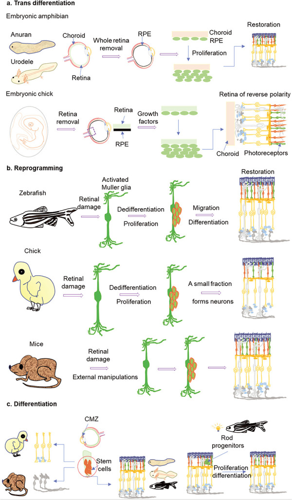
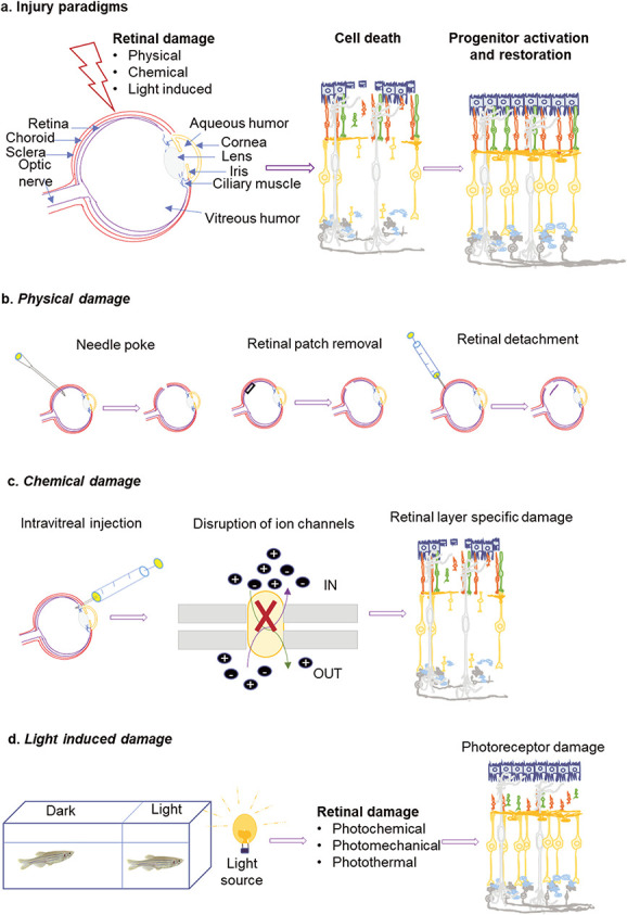
Unlike mammals, vertebrates such as fishes and frogs exhibit remarkable tissue regeneration including the central nervous system. Retina being part of the central nervous system has attracted the interest of several research groups to explore its regenerative ability in different vertebrate models including mice. Fishes and frogs completely restore the size, shape and tissue structure of an injured retina. Several studies have unraveled molecular mechanisms underlying retina regeneration. In teleosts, soon after injury, the Müller glial cells of the retina reprogram to form a proliferating population of Müller glia-derived progenitor cells capable of differentiating into various neural cell types and Müller glia. In amphibians, the transdifferentiation of retinal pigment epithelium and differentiation of ciliary marginal zone cells contribute to retina regeneration. In chicks and mice, supplementation with external growth factors or genetic modifications cause a partial regenerative response in the damaged retina. The initiation of retina regeneration is achieved through sequential orchestration of gene expression through controlled modulations in the genetic and epigenetic landscape of the progenitor cells. Several developmental biology pathways are turned on during the Müller glia reprogramming, retinal pigment epithelium transdifferentiation and ciliary marginal zone differentiation. Further, several tumorigenic pathways and gene expression events also contribute to the complete regeneration cascade of events. In this review, we address the various retinal injury paradigms and subsequent gene expression events governed in different vertebrate species. Further, we compared how vertebrates such as teleost fishes and amphibians can achieve excellent regenerative responses in the retina compared with their mammalian counterparts.

 求助内容:
求助内容: 应助结果提醒方式:


