{"title":"Erb-(IL10)2 ameliorates radiation-induced skin injury through eliminate oxygen free radicals.","authors":"Jiahe Xu, Jiaxing Zhu, Qi Zhao, Jiao Xue, Songbing Qin","doi":"10.1002/pro6.1193","DOIUrl":null,"url":null,"abstract":"<p><strong>Objective: </strong>Radiation-induced skin injury (RISI) remains a serious concern during radiotherapy. IL-10 is considered as an immune suppressive cytokine by inhibiting the secretion of the proinflammatory cytokines in cells. The aim of this study was to evaluate the protective role of Erb (IL10) 2 against ionizing radiation.</p><p><strong>Methods: </strong>We fused Interleukin 10 (IL-10) dimer onto an anti-epidermal growth factor receptor antibody Cetuximab (Erbitux) to form a new bispecific protein Erb-(IL10)2. The protective effect and biological activity of Erb-(IL10)2 was measured in model of RISI.</p><p><strong>Results: </strong>Under the condition of 20 Gy irradiation, surviving cells in the IR group decreased significantly compared with the non-IR group (<i>p</i> = 0.0021). The survival rates of HaCaT (<i>p</i> = 0.0038) and WS1 (<i>p</i> = 0.0003) cells were significantly increased after IL-10 treatment. The apoptosis rates of HaCaT (<i>p</i> = 0.0048) and WS1 (<i>p</i> = 0.0074) cells in the IL-10 group were significantly lower compared to the NC group. Under 20 Gy irradiation conditions, IL-10 fusion protein reduced the level of reactive oxygen in HaCaT (<i>p</i> = 0.0046) and WS1 (<i>p</i><0.0001) cells compared to the control group. Relatively normal granular mitochondrial morphology was observed in the IL-10 group after 20 Gy X-ray irradiation compared with the NC group. Ater 35 Gy electron radiation, the levels of reactive oxygen species in the skin tissue of C57/B6 mice injected with IL-10 fusion protein were significantly lower than those in the PBS group (<i>p</i> = 0.001). Compared with the PBS group and the other IL-10 groups, the group treated with 0.2 mg/kg IL-10 showed a significant decrease in MDA level (<i>p</i> = 0.0024). Compared with the PBS group, the thickness of the stratum corneum in groups treated with 0.05, 0.1 and 0.2 mg/kg IL-10 decreased, and the skin appendages were well-preserved. In the group treated with 0.2 mg/kg IL-10, the skin tissue structure was still relatively intact, and the masson staining area was smaller than that of the PBS group.</p><p><strong>Conclusion: </strong>IL-10 plays a role in inhibiting radioactive fibrosis in radioactive skin injury. IL-10 has a protective effect on skin cell damage after ionizing radiation irradiation both in vitro and in vivo. Moreover, IL-10 plays a role in inhibiting radioactive fibrosis in radioactive skin injury.</p>","PeriodicalId":32406,"journal":{"name":"Precision Radiation Oncology","volume":"7 1","pages":"92-100"},"PeriodicalIF":2.1000,"publicationDate":"2023-06-12","publicationTypes":"Journal Article","fieldsOfStudy":null,"isOpenAccess":false,"openAccessPdf":"https://www.ncbi.nlm.nih.gov/pmc/articles/PMC11935052/pdf/","citationCount":"0","resultStr":null,"platform":"Semanticscholar","paperid":null,"PeriodicalName":"Precision Radiation Oncology","FirstCategoryId":"1085","ListUrlMain":"https://doi.org/10.1002/pro6.1193","RegionNum":0,"RegionCategory":null,"ArticlePicture":[],"TitleCN":null,"AbstractTextCN":null,"PMCID":null,"EPubDate":"2023/6/1 0:00:00","PubModel":"eCollection","JCR":"Q4","JCRName":"Medicine","Score":null,"Total":0}
引用次数: 0
Abstract
Objective: Radiation-induced skin injury (RISI) remains a serious concern during radiotherapy. IL-10 is considered as an immune suppressive cytokine by inhibiting the secretion of the proinflammatory cytokines in cells. The aim of this study was to evaluate the protective role of Erb (IL10) 2 against ionizing radiation.
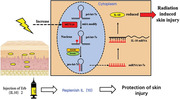
Methods: We fused Interleukin 10 (IL-10) dimer onto an anti-epidermal growth factor receptor antibody Cetuximab (Erbitux) to form a new bispecific protein Erb-(IL10)2. The protective effect and biological activity of Erb-(IL10)2 was measured in model of RISI.
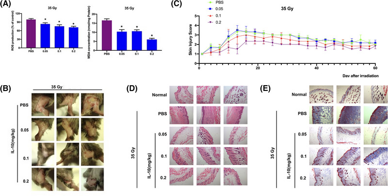
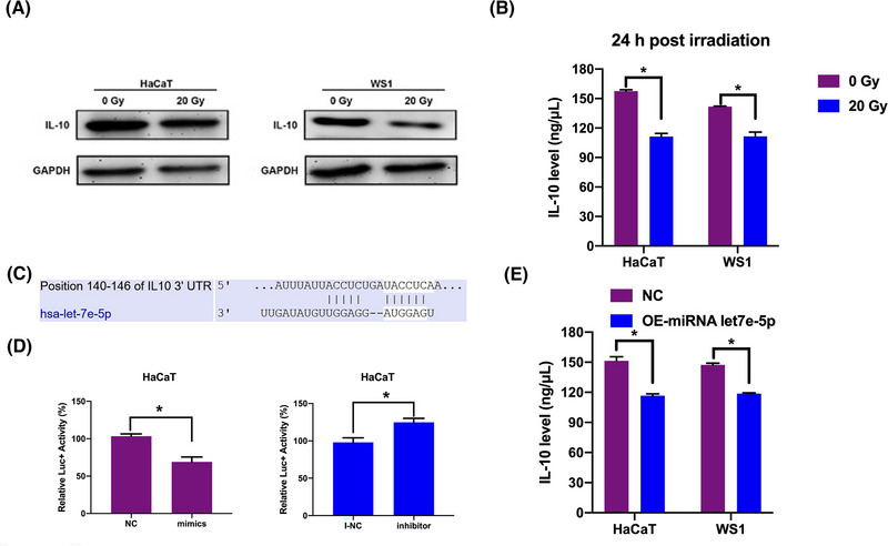
Results: Under the condition of 20 Gy irradiation, surviving cells in the IR group decreased significantly compared with the non-IR group (p = 0.0021). The survival rates of HaCaT (p = 0.0038) and WS1 (p = 0.0003) cells were significantly increased after IL-10 treatment. The apoptosis rates of HaCaT (p = 0.0048) and WS1 (p = 0.0074) cells in the IL-10 group were significantly lower compared to the NC group. Under 20 Gy irradiation conditions, IL-10 fusion protein reduced the level of reactive oxygen in HaCaT (p = 0.0046) and WS1 (p<0.0001) cells compared to the control group. Relatively normal granular mitochondrial morphology was observed in the IL-10 group after 20 Gy X-ray irradiation compared with the NC group. Ater 35 Gy electron radiation, the levels of reactive oxygen species in the skin tissue of C57/B6 mice injected with IL-10 fusion protein were significantly lower than those in the PBS group (p = 0.001). Compared with the PBS group and the other IL-10 groups, the group treated with 0.2 mg/kg IL-10 showed a significant decrease in MDA level (p = 0.0024). Compared with the PBS group, the thickness of the stratum corneum in groups treated with 0.05, 0.1 and 0.2 mg/kg IL-10 decreased, and the skin appendages were well-preserved. In the group treated with 0.2 mg/kg IL-10, the skin tissue structure was still relatively intact, and the masson staining area was smaller than that of the PBS group.
Conclusion: IL-10 plays a role in inhibiting radioactive fibrosis in radioactive skin injury. IL-10 has a protective effect on skin cell damage after ionizing radiation irradiation both in vitro and in vivo. Moreover, IL-10 plays a role in inhibiting radioactive fibrosis in radioactive skin injury.

 求助内容:
求助内容: 应助结果提醒方式:


