{"title":"Quantification of vascular morphology in optical coherence tomography angiography in primary open angle glaucoma","authors":"Praneeth Kalva , Rubeel Akram , Priya Mekala , Monica Patel , Sruthi Suresh , Karanjit S. Kooner","doi":"10.1016/j.aopr.2023.05.002","DOIUrl":null,"url":null,"abstract":"<div><h3>Purpose</h3><p>To quantitatively measure and compare the vascular morphology in healthy eyes and eyes with primary open-angle glaucoma (POAG) using optical coherence tomography angiography (OCTA) scans.</p></div><div><h3>Methods</h3><p>This is a retrospective and cross-sectional study which include healthy individuals and individuals with POAG that underwent OCTA imaging at an academic center's glaucoma clinic. We analyzed OCTA scans of the macula and optic nerve head (ONH) of one eye from each subject to quantitatively measure vessel density (VD), vessel length density (VLD), and branchpoint density (BPD). We compared these 3 parameters between the healthy and POAG groups and used logistic regression classification models to determine their diagnostic value in differentiating healthy and glaucomatous eyes.</p></div><div><h3>Results</h3><p>We included 49 healthy subjects and 49 subjects with POAG. After age-adjusted analysis, the parameters of VD, VLD, and BPD were significantly reduced in eyes with POAG (<em>P</em> < 0.001) in all scan layers and most significantly around the ONH. The parameter with the best performances were radial peripapillary capillary (RPC) VD [AUC (areas under the curve): 0.939 (0.891, 0.987)] which had statistically higher performances (<em>P</em> < 0.05) than parameters in the superficial or deep layers. All 3 parameters in the RPC layer had statistically similar performances.</p></div><div><h3>Conclusions</h3><p>We found that VD, VLD, and BPD were reduced in glaucomatous eyes. The morphologic parameters of VLD and BPD had similar performances to the traditional parameter of VD in RPC layers. Our results suggest that vascular morphology parameters may provide additional value in the diagnosis and evaluation of glaucoma.</p></div>","PeriodicalId":72103,"journal":{"name":"Advances in ophthalmology practice and research","volume":"3 3","pages":"Pages 119-125"},"PeriodicalIF":3.4000,"publicationDate":"2023-08-01","publicationTypes":"Journal Article","fieldsOfStudy":null,"isOpenAccess":false,"openAccessPdf":"https://ftp.ncbi.nlm.nih.gov/pub/pmc/oa_pdf/47/10/main.PMC10577834.pdf","citationCount":"0","resultStr":null,"platform":"Semanticscholar","paperid":null,"PeriodicalName":"Advances in ophthalmology practice and research","FirstCategoryId":"1085","ListUrlMain":"https://www.sciencedirect.com/science/article/pii/S266737622300015X","RegionNum":0,"RegionCategory":null,"ArticlePicture":[],"TitleCN":null,"AbstractTextCN":null,"PMCID":null,"EPubDate":"","PubModel":"","JCR":"","JCRName":"","Score":null,"Total":0}
引用次数: 0
Abstract
Purpose
To quantitatively measure and compare the vascular morphology in healthy eyes and eyes with primary open-angle glaucoma (POAG) using optical coherence tomography angiography (OCTA) scans.
Methods
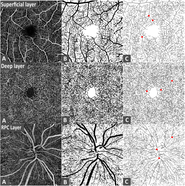
This is a retrospective and cross-sectional study which include healthy individuals and individuals with POAG that underwent OCTA imaging at an academic center's glaucoma clinic. We analyzed OCTA scans of the macula and optic nerve head (ONH) of one eye from each subject to quantitatively measure vessel density (VD), vessel length density (VLD), and branchpoint density (BPD). We compared these 3 parameters between the healthy and POAG groups and used logistic regression classification models to determine their diagnostic value in differentiating healthy and glaucomatous eyes.
Results
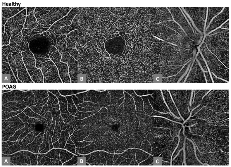
We included 49 healthy subjects and 49 subjects with POAG. After age-adjusted analysis, the parameters of VD, VLD, and BPD were significantly reduced in eyes with POAG (P < 0.001) in all scan layers and most significantly around the ONH. The parameter with the best performances were radial peripapillary capillary (RPC) VD [AUC (areas under the curve): 0.939 (0.891, 0.987)] which had statistically higher performances (P < 0.05) than parameters in the superficial or deep layers. All 3 parameters in the RPC layer had statistically similar performances.
Conclusions
We found that VD, VLD, and BPD were reduced in glaucomatous eyes. The morphologic parameters of VLD and BPD had similar performances to the traditional parameter of VD in RPC layers. Our results suggest that vascular morphology parameters may provide additional value in the diagnosis and evaluation of glaucoma.

 求助内容:
求助内容: 应助结果提醒方式:


