Sara B Hobday, Leila J Mady, Alec M Jacobson, Christopher H Rassekh
{"title":"CIC-DUX4 Sarcoma Involving the Skull Base: A Rare Presentation and Review of the Literature.","authors":"Sara B Hobday, Leila J Mady, Alec M Jacobson, Christopher H Rassekh","doi":"10.1055/a-2166-5688","DOIUrl":null,"url":null,"abstract":"<p><p><b>Background</b> CIC-DUX4 sarcoma is a rare, aggressive tumor that is difficult to diagnose. Although it is closely related to Ewing's sarcoma, each is a distinct pathologic entity and both have been previously reported in the skin, lymph nodes, and viscera. We report the first description of CIC-DUX4 involving the posterior cranial fossa and review the distinctive symptomatology, morphology, immunoprofile, and genetic signature that differentiate this rare tumor. <b>Case Report</b> A 32-year-old man presented with an enlarging right lateral neck mass, progressive hoarseness, and orofacial pain. Biopsy revealed a high-grade undifferentiated malignant neoplasm. Imaging demonstrated an 8-cm mass in the right neck extending to the skull base and abutting the carotid sheath, in addition to pulmonary nodules and pelvic lymphadenopathy. Despite initial response to chemotherapy, he experienced disease progression and underwent surgical resection, radical neck dissection, and brachytherapy. Definitive pathologic diagnosis was achieved with next-generation sequencing. Within weeks of treatment, he developed symptoms reflecting progression of disease involving the neck, posterior cranial fossa, and lung. Adjuvant chemotherapy was planned, but the patient succumbed to his disease prior to initiation of further therapy. <b>Conclusion</b> CIC-DUX4 sarcomas are uncommon and can progress rapidly. Diagnosis requires either fluorescence in situ hybridization or next-generation sequencing. Due to its rarity, there is no standard-of-care treatment for this tumor and further investigations are needed to understand disease behavior and develop targeted therapeutic modalities.</p>","PeriodicalId":44256,"journal":{"name":"Journal of Neurological Surgery Reports","volume":"84 4","pages":"e124-e128"},"PeriodicalIF":0.7000,"publicationDate":"2023-10-13","publicationTypes":"Journal Article","fieldsOfStudy":null,"isOpenAccess":false,"openAccessPdf":"https://ftp.ncbi.nlm.nih.gov/pub/pmc/oa_pdf/62/73/10-1055-a-2166-5688.PMC10575739.pdf","citationCount":"0","resultStr":null,"platform":"Semanticscholar","paperid":null,"PeriodicalName":"Journal of Neurological Surgery Reports","FirstCategoryId":"1085","ListUrlMain":"https://doi.org/10.1055/a-2166-5688","RegionNum":0,"RegionCategory":null,"ArticlePicture":[],"TitleCN":null,"AbstractTextCN":null,"PMCID":null,"EPubDate":"2023/10/1 0:00:00","PubModel":"eCollection","JCR":"Q4","JCRName":"CLINICAL NEUROLOGY","Score":null,"Total":0}
引用次数: 0
Abstract
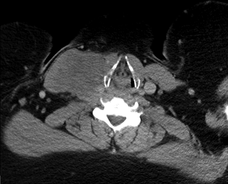
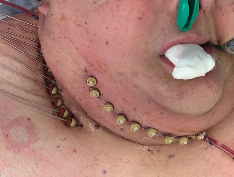
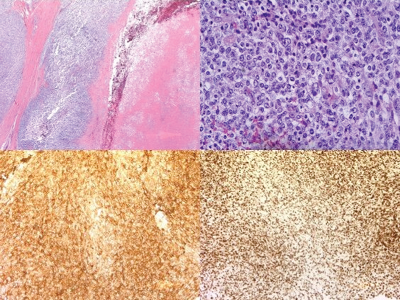
Background CIC-DUX4 sarcoma is a rare, aggressive tumor that is difficult to diagnose. Although it is closely related to Ewing's sarcoma, each is a distinct pathologic entity and both have been previously reported in the skin, lymph nodes, and viscera. We report the first description of CIC-DUX4 involving the posterior cranial fossa and review the distinctive symptomatology, morphology, immunoprofile, and genetic signature that differentiate this rare tumor. Case Report A 32-year-old man presented with an enlarging right lateral neck mass, progressive hoarseness, and orofacial pain. Biopsy revealed a high-grade undifferentiated malignant neoplasm. Imaging demonstrated an 8-cm mass in the right neck extending to the skull base and abutting the carotid sheath, in addition to pulmonary nodules and pelvic lymphadenopathy. Despite initial response to chemotherapy, he experienced disease progression and underwent surgical resection, radical neck dissection, and brachytherapy. Definitive pathologic diagnosis was achieved with next-generation sequencing. Within weeks of treatment, he developed symptoms reflecting progression of disease involving the neck, posterior cranial fossa, and lung. Adjuvant chemotherapy was planned, but the patient succumbed to his disease prior to initiation of further therapy. Conclusion CIC-DUX4 sarcomas are uncommon and can progress rapidly. Diagnosis requires either fluorescence in situ hybridization or next-generation sequencing. Due to its rarity, there is no standard-of-care treatment for this tumor and further investigations are needed to understand disease behavior and develop targeted therapeutic modalities.

 求助内容:
求助内容: 应助结果提醒方式:


