{"title":"Difference in arterial FDG accumulation in healthy study participants between digital PET/CT and standard PET/CT.","authors":"Noriko Nitta, Rika Yoshimatsu, Hitomi Iwasa, Kousuke Nakaji, Kana Miyatake, Miki Nishimori, Tomohiro Matsumoto, Tomoaki Yamanishi, Takuji Yamagami","doi":"10.1007/s12149-023-01875-4","DOIUrl":null,"url":null,"abstract":"<p><strong>Objective: </strong>To evaluate the differences in FDG accumulation in arteries throughout the body between digital and standard PET/CT.</p><p><strong>Methods: </strong>Forty-six people who had FDG-PET examinations with a digital PET/CT scanner for health screening between April 2020 and March 2021 and had previous examinations with a standard PET/CT scanner were the study participants. FDG accumulation in arteries throughout the body was visually assessed in each segment. Scan was considered positive when arterial FDG accumulation was equal to or greater than that of the liver. The positivity rates for general arteries and each arterial segment were compared between the two kinds of scanners. If any one of the arterial segments was considered positive, the general arteries were classified as positive. Moreover, the rate of change in results from the standard PET/CT to the digital scanner in the same individual (negative to positive, positive to negative) was examined.</p><p><strong>Results: </strong>In the evaluation of general arteries, the positivity rates were 21.7% (10 cases) for the standard PET/CT, whereas positive rates were 97.8% (45 cases) for the digital PET/CT (p < 0.001). In all arterial segments, the positivity rate was significantly higher with the digital PET/CT compared to the standard PET/CT; those with the digital PET/CT were, respectively, 95.7%, 87.0%, 73.9%, 37.0%, 34.8%, and 21.7% in the femoral, brachial, aortic, subclavian, iliac, and carotid arteries. On the other hand, those with the standard PET/CT were 13.0%, 13.0%, 19.6%, 2.2%, 0%, and 4.4% in segments in the above order. Changes from negative to positive were shown in many cases; 35 cases (76.0%) of general arteries, 38 cases (82.6%) for the femoral artery, and 34 cases (73.9%) for the brachial artery. The exception was one case in which a change from positive to negative was confirmed in the carotid artery. In all arteries considered to be positive, FDG accumulation was not greater than but was equal to that in the liver with both scanners.</p><p><strong>Conclusions: </strong>Arterial FDG accumulation was significantly higher with digital PET/CT compared to conventional PET/CT. With digital PET/CT, an arterial FDG accumulation equal to the liver may not to be considered as abnormal accumulation.</p>","PeriodicalId":8007,"journal":{"name":"Annals of Nuclear Medicine","volume":null,"pages":null},"PeriodicalIF":2.5000,"publicationDate":"2024-02-01","publicationTypes":"Journal Article","fieldsOfStudy":null,"isOpenAccess":false,"openAccessPdf":"","citationCount":"0","resultStr":null,"platform":"Semanticscholar","paperid":null,"PeriodicalName":"Annals of Nuclear Medicine","FirstCategoryId":"3","ListUrlMain":"https://doi.org/10.1007/s12149-023-01875-4","RegionNum":4,"RegionCategory":"医学","ArticlePicture":[],"TitleCN":null,"AbstractTextCN":null,"PMCID":null,"EPubDate":"2023/10/18 0:00:00","PubModel":"Epub","JCR":"Q2","JCRName":"RADIOLOGY, NUCLEAR MEDICINE & MEDICAL IMAGING","Score":null,"Total":0}
引用次数: 0
Abstract
Objective: To evaluate the differences in FDG accumulation in arteries throughout the body between digital and standard PET/CT.
Methods: Forty-six people who had FDG-PET examinations with a digital PET/CT scanner for health screening between April 2020 and March 2021 and had previous examinations with a standard PET/CT scanner were the study participants. FDG accumulation in arteries throughout the body was visually assessed in each segment. Scan was considered positive when arterial FDG accumulation was equal to or greater than that of the liver. The positivity rates for general arteries and each arterial segment were compared between the two kinds of scanners. If any one of the arterial segments was considered positive, the general arteries were classified as positive. Moreover, the rate of change in results from the standard PET/CT to the digital scanner in the same individual (negative to positive, positive to negative) was examined.
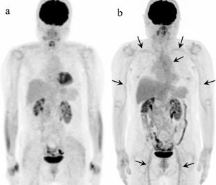
Results: In the evaluation of general arteries, the positivity rates were 21.7% (10 cases) for the standard PET/CT, whereas positive rates were 97.8% (45 cases) for the digital PET/CT (p < 0.001). In all arterial segments, the positivity rate was significantly higher with the digital PET/CT compared to the standard PET/CT; those with the digital PET/CT were, respectively, 95.7%, 87.0%, 73.9%, 37.0%, 34.8%, and 21.7% in the femoral, brachial, aortic, subclavian, iliac, and carotid arteries. On the other hand, those with the standard PET/CT were 13.0%, 13.0%, 19.6%, 2.2%, 0%, and 4.4% in segments in the above order. Changes from negative to positive were shown in many cases; 35 cases (76.0%) of general arteries, 38 cases (82.6%) for the femoral artery, and 34 cases (73.9%) for the brachial artery. The exception was one case in which a change from positive to negative was confirmed in the carotid artery. In all arteries considered to be positive, FDG accumulation was not greater than but was equal to that in the liver with both scanners.
Conclusions: Arterial FDG accumulation was significantly higher with digital PET/CT compared to conventional PET/CT. With digital PET/CT, an arterial FDG accumulation equal to the liver may not to be considered as abnormal accumulation.
期刊介绍:
Annals of Nuclear Medicine is an official journal of the Japanese Society of Nuclear Medicine. It develops the appropriate application of radioactive substances and stable nuclides in the field of medicine.
The journal promotes the exchange of ideas and information and research in nuclear medicine and includes the medical application of radionuclides and related subjects. It presents original articles, short communications, reviews and letters to the editor.

 求助内容:
求助内容: 应助结果提醒方式:


