Ryley Mancine, Paul Kowalski, William McMillan, Nicole Geske, Loro Kujjo
{"title":"Discovering Pathologies in the Anatomy Lab: The Case of Brachial Plexopathy Mimicking Neurological Thoracic Outlet Syndrome.","authors":"Ryley Mancine, Paul Kowalski, William McMillan, Nicole Geske, Loro Kujjo","doi":"","DOIUrl":null,"url":null,"abstract":"<p><strong>Context: </strong>Well-established human anatomy labs with access to expert faculty are exceedingly valuable tools to medical student education. In this manuscript, we detail an infero-lateral subclavicular lipoma which was discovered as a result of the utilization of both those labs and expert faculty. This lipoma may have caused brachial plexopathy or may serve as an unusual cause of neurologic thoracic outlet syndrome (NTOS) due to the location of the mass.</p><p><strong>Educational case presentation: </strong>During prosection of a donor in the human anatomy lab, a mass was discovered by a medical student. This medical student utilized the human anatomy lab faculty members and resources to identify the mass as a lipoma. The lipoma compressed the lateral cord of the brachial plexus and the suprascapular nerve, but no diagnosis of NTOS or brachial plexopathy was made during the life of the donor, nor was any surgical intervention indicated. Removal of the lipoma immediately relieved stress upon the nerves. Histochemical analysis confirmed the diagnosis of a lipoma and demonstrated almost only mature adipocytes.</p><p><strong>Conclusion: </strong>The authors concluded that the lipoma of this patient was not identifiable with computerized tomography imaging modalities, despite ultrasound demonstrating a hyperechoic outline of the mass in the cadaver of the patient. It is very likely that this lipoma had not been diagnosed previously due to the atypical location of the tumor. Equally, typical surgical methods associated with brachial plexopathy or NTOS treatment would be difficult or more complicated, due to the lateral and inferior location of the lipoma. Physicians treating thoracic outlet syndrome-type symptoms without resolution should consider potential non-malignant obstruction located outside the thoracic outlet, toward the extremity. Deep palpatory methods and physical therapy should be considered until diagnosis is certain, as ultrasound would be difficult and typical transaxillary surgical methods would be nonhelpful. Medical students and early-career residents and physicians should be aware of the resources provided to them via campus human anatomy laboratories which they may utilize to further their understanding and knowledge of specific pathologies.</p>","PeriodicalId":74853,"journal":{"name":"Spartan medical research journal","volume":"5 2","pages":"14179"},"PeriodicalIF":0.0000,"publicationDate":"2020-10-30","publicationTypes":"Journal Article","fieldsOfStudy":null,"isOpenAccess":false,"openAccessPdf":"https://www.ncbi.nlm.nih.gov/pmc/articles/PMC7746061/pdf/","citationCount":"0","resultStr":null,"platform":"Semanticscholar","paperid":null,"PeriodicalName":"Spartan medical research journal","FirstCategoryId":"1085","ListUrlMain":"","RegionNum":0,"RegionCategory":null,"ArticlePicture":[],"TitleCN":null,"AbstractTextCN":null,"PMCID":null,"EPubDate":"","PubModel":"","JCR":"","JCRName":"","Score":null,"Total":0}
引用次数: 0
Abstract
Context: Well-established human anatomy labs with access to expert faculty are exceedingly valuable tools to medical student education. In this manuscript, we detail an infero-lateral subclavicular lipoma which was discovered as a result of the utilization of both those labs and expert faculty. This lipoma may have caused brachial plexopathy or may serve as an unusual cause of neurologic thoracic outlet syndrome (NTOS) due to the location of the mass.
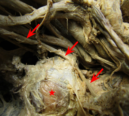
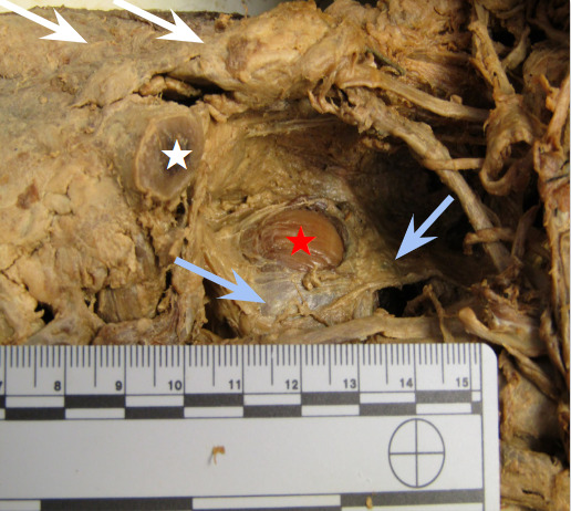
Educational case presentation: During prosection of a donor in the human anatomy lab, a mass was discovered by a medical student. This medical student utilized the human anatomy lab faculty members and resources to identify the mass as a lipoma. The lipoma compressed the lateral cord of the brachial plexus and the suprascapular nerve, but no diagnosis of NTOS or brachial plexopathy was made during the life of the donor, nor was any surgical intervention indicated. Removal of the lipoma immediately relieved stress upon the nerves. Histochemical analysis confirmed the diagnosis of a lipoma and demonstrated almost only mature adipocytes.
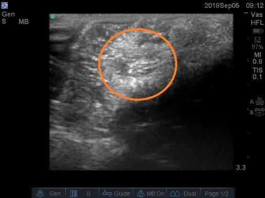
Conclusion: The authors concluded that the lipoma of this patient was not identifiable with computerized tomography imaging modalities, despite ultrasound demonstrating a hyperechoic outline of the mass in the cadaver of the patient. It is very likely that this lipoma had not been diagnosed previously due to the atypical location of the tumor. Equally, typical surgical methods associated with brachial plexopathy or NTOS treatment would be difficult or more complicated, due to the lateral and inferior location of the lipoma. Physicians treating thoracic outlet syndrome-type symptoms without resolution should consider potential non-malignant obstruction located outside the thoracic outlet, toward the extremity. Deep palpatory methods and physical therapy should be considered until diagnosis is certain, as ultrasound would be difficult and typical transaxillary surgical methods would be nonhelpful. Medical students and early-career residents and physicians should be aware of the resources provided to them via campus human anatomy laboratories which they may utilize to further their understanding and knowledge of specific pathologies.

 求助内容:
求助内容: 应助结果提醒方式:


