{"title":"Evaluation of the relationship between sphenoid sinus morphology and area and volume by computed tomography.","authors":"Mehmet Serindere, Ceren Aktuna Belgin","doi":"10.1007/s11282-023-00711-9","DOIUrl":null,"url":null,"abstract":"<p><strong>Objectives: </strong>The aim of this study is to investigate the effect of sphenoid sinus pneumatization types, Onodi cell (OC), internal carotid artery (ICA), optic nerve (ON) on sinus volume and area on computed tomography (CT) images.</p><p><strong>Methods: </strong>The CT images of 150 patients were evaluated. Sphenoid sinus pneumatization types, OC prevalence, protrusion and dehiscence of ICA and ON, the volume and area were evaluated.</p><p><strong>Results: </strong>The sinus volume and area were statistically higher in patients with bilateral protrusion of ICA and ON then patients without protrusion of ICA and ON. The mean volume and area of sinus were 9949.4 ± 351.0 mm<sup>3</sup> and 4570.9 ± 1604.9 mm<sup>2</sup>, respectively. The volume and area of sphenoid sinus did not differ significantly between groups with and without OC. The postsellar b type sphenoid sinus had the highest volume, while conchal type has the least volume.</p><p><strong>Conclusions: </strong>Bilateral protrusion and dehiscence of ICA and bilateral protrusion of ON caused a significant increase in the sphenoid sinus volume and area. The presence of ICA and ON, the pneumatization of the sinus is an anatomical structure that can affect the sinus volume and area. Before the operation, three-dimensional evaluation should be performed to determine whether these structures are bilateral/unilateral and it should be remembered that the sinus volume and area can change.</p>","PeriodicalId":56103,"journal":{"name":"Oral Radiology","volume":" ","pages":"138-147"},"PeriodicalIF":1.6000,"publicationDate":"2024-04-01","publicationTypes":"Journal Article","fieldsOfStudy":null,"isOpenAccess":false,"openAccessPdf":"","citationCount":"0","resultStr":null,"platform":"Semanticscholar","paperid":null,"PeriodicalName":"Oral Radiology","FirstCategoryId":"3","ListUrlMain":"https://doi.org/10.1007/s11282-023-00711-9","RegionNum":3,"RegionCategory":"医学","ArticlePicture":[],"TitleCN":null,"AbstractTextCN":null,"PMCID":null,"EPubDate":"2023/9/25 0:00:00","PubModel":"Epub","JCR":"Q3","JCRName":"DENTISTRY, ORAL SURGERY & MEDICINE","Score":null,"Total":0}
引用次数: 0
Abstract
Objectives: The aim of this study is to investigate the effect of sphenoid sinus pneumatization types, Onodi cell (OC), internal carotid artery (ICA), optic nerve (ON) on sinus volume and area on computed tomography (CT) images.
Methods: The CT images of 150 patients were evaluated. Sphenoid sinus pneumatization types, OC prevalence, protrusion and dehiscence of ICA and ON, the volume and area were evaluated.
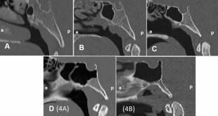
Results: The sinus volume and area were statistically higher in patients with bilateral protrusion of ICA and ON then patients without protrusion of ICA and ON. The mean volume and area of sinus were 9949.4 ± 351.0 mm3 and 4570.9 ± 1604.9 mm2, respectively. The volume and area of sphenoid sinus did not differ significantly between groups with and without OC. The postsellar b type sphenoid sinus had the highest volume, while conchal type has the least volume.
Conclusions: Bilateral protrusion and dehiscence of ICA and bilateral protrusion of ON caused a significant increase in the sphenoid sinus volume and area. The presence of ICA and ON, the pneumatization of the sinus is an anatomical structure that can affect the sinus volume and area. Before the operation, three-dimensional evaluation should be performed to determine whether these structures are bilateral/unilateral and it should be remembered that the sinus volume and area can change.
期刊介绍:
As the official English-language journal of the Japanese Society for Oral and Maxillofacial Radiology and the Asian Academy of Oral and Maxillofacial Radiology, Oral Radiology is intended to be a forum for international collaboration in head and neck diagnostic imaging and all related fields. Oral Radiology features cutting-edge research papers, review articles, case reports, and technical notes from both the clinical and experimental fields. As membership in the Society is not a prerequisite, contributions are welcome from researchers and clinicians worldwide.

 求助内容:
求助内容: 应助结果提醒方式:


