Rawa Bapir, Ismaeel Aghaways, Rawa M Ali, Saman S Fakhralddin, Rebaz M Ali, Rezheen J Rashid, Ari M Abdullah, Muhammed Bag A Ali, Karukh K Mohammed, Hiwa O Abdullah, Fahmi H Kakamad
{"title":"Spermatocytic tumor of the testis: A case report and mini‑review of the literature.","authors":"Rawa Bapir, Ismaeel Aghaways, Rawa M Ali, Saman S Fakhralddin, Rebaz M Ali, Rezheen J Rashid, Ari M Abdullah, Muhammed Bag A Ali, Karukh K Mohammed, Hiwa O Abdullah, Fahmi H Kakamad","doi":"10.3892/mi.2023.111","DOIUrl":null,"url":null,"abstract":"<p><p>Spermatocytic tumors are a rare type of testicular cancer, comprising <1% of all testicular malignancies. This type of cancer typically affects males in their 60s and 70s and rarely metastasizes; however, it poses a threat to the health of affected individuals if left untreated. The present study describes the case of a 68-year-old male patient with this type of tumor, including a presentation of his initial symptoms, treatment and subsequent monitoring. A male patient, aged 68 years, visited the authors' clinic with an asymptomatic mass in the right testicle. The mass had been progressively increasing in size for a duration of 5 years following a history of blunt injury. During the examination, a noticeable, painless enlargement was detected in the right testis, whereas the left testis appeared to be in a normal state. Tumor markers were within normal limits. Imaging revealed a complex mass (11x8x7 cm) almost replacing the right testis, with no detectable lymph nodes. A right radical orchidectomy was performed under spinal anesthesia. A histopathological examination revealed a spermatocytic tumor. The post-operative period was uneventful, with no metastasis detected in the CT scans. The patient was discharged with instructions for regular follow-up appointments. The case presented herein highlights a rare spermatocytic tumor in a 68-year-old male. The early detection and treatment of testicular tumors, regardless of age, are crucial for a good prognosis.</p>","PeriodicalId":74161,"journal":{"name":"Medicine international","volume":"3 5","pages":"51"},"PeriodicalIF":0.0000,"publicationDate":"2023-09-11","publicationTypes":"Journal Article","fieldsOfStudy":null,"isOpenAccess":false,"openAccessPdf":"https://www.ncbi.nlm.nih.gov/pmc/articles/PMC10557091/pdf/","citationCount":"0","resultStr":null,"platform":"Semanticscholar","paperid":null,"PeriodicalName":"Medicine international","FirstCategoryId":"1085","ListUrlMain":"https://doi.org/10.3892/mi.2023.111","RegionNum":0,"RegionCategory":null,"ArticlePicture":[],"TitleCN":null,"AbstractTextCN":null,"PMCID":null,"EPubDate":"2023/9/1 0:00:00","PubModel":"eCollection","JCR":"","JCRName":"","Score":null,"Total":0}
引用次数: 0
Abstract
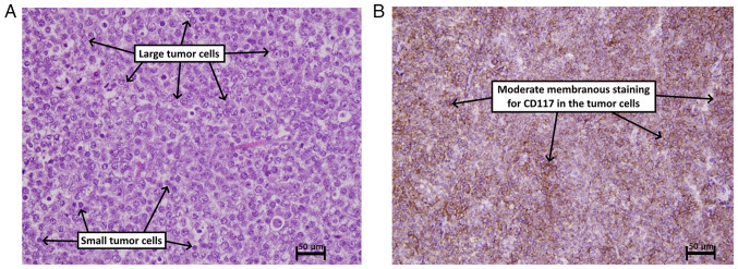
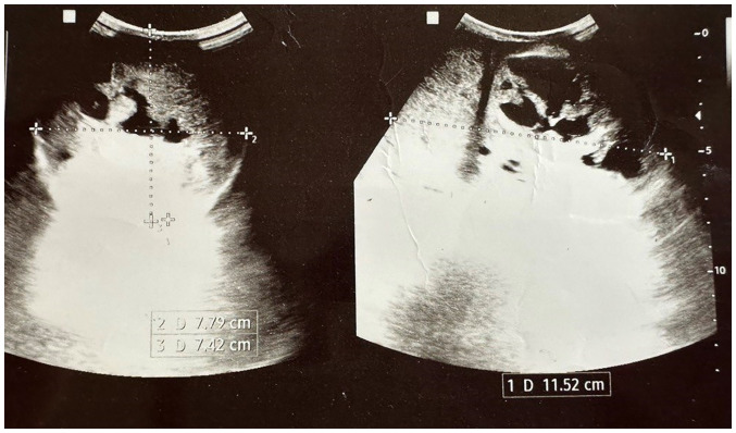
Spermatocytic tumors are a rare type of testicular cancer, comprising <1% of all testicular malignancies. This type of cancer typically affects males in their 60s and 70s and rarely metastasizes; however, it poses a threat to the health of affected individuals if left untreated. The present study describes the case of a 68-year-old male patient with this type of tumor, including a presentation of his initial symptoms, treatment and subsequent monitoring. A male patient, aged 68 years, visited the authors' clinic with an asymptomatic mass in the right testicle. The mass had been progressively increasing in size for a duration of 5 years following a history of blunt injury. During the examination, a noticeable, painless enlargement was detected in the right testis, whereas the left testis appeared to be in a normal state. Tumor markers were within normal limits. Imaging revealed a complex mass (11x8x7 cm) almost replacing the right testis, with no detectable lymph nodes. A right radical orchidectomy was performed under spinal anesthesia. A histopathological examination revealed a spermatocytic tumor. The post-operative period was uneventful, with no metastasis detected in the CT scans. The patient was discharged with instructions for regular follow-up appointments. The case presented herein highlights a rare spermatocytic tumor in a 68-year-old male. The early detection and treatment of testicular tumors, regardless of age, are crucial for a good prognosis.

 求助内容:
求助内容: 应助结果提醒方式:


