Peng-Bo Zhu, Yeon-Dong Kim, Ha Yeong Jeong, Miyoung Yang, Hyung-Sun Won
{"title":"New insight into the mandibular nerve at the foramen ovale level for percutaneous radiofrequency thermocoagulation.","authors":"Peng-Bo Zhu, Yeon-Dong Kim, Ha Yeong Jeong, Miyoung Yang, Hyung-Sun Won","doi":"10.3344/kjp.23186","DOIUrl":null,"url":null,"abstract":"<p><strong>Background: </strong>Percutaneous radiofrequency thermocoagulation (RFTC) has been widely utilized in the management of trigeminal neuralgia. Despite using image guidance, accurate needle positioning into the target area still remains a critical element for achieving a successful outcome. This study was performed to precisely clarify the anatomical information required to ensure that the electrode tip is placed on the sensory component of the mandibular nerve (MN) at the foramen ovale (FO) level.</p><p><strong>Methods: </strong>The study used 50 hemi-half heads from 26 South Korean adult cadavers.</p><p><strong>Results: </strong>The cross-sectioned anterior and posterior divisions of the MN at the FO level could be distinguished based on an irregular boundary and color difference. The anterior division was clearly brighter than the posterior one. The anterior division of the MN at the FO level was located at the whole anterior (38.0%), anteromedial (6.0%), anterior center (8.0%), and anterolateral (22.0%) parts. The posterior division was often located at the whole posterior or posterolateral parts of the MN at the FO level. The anterior divisions covered the whole MN except for the medial half of the posterolateral part in the overwrapped images of the cross-sectional areas of the MN at the FO level. The cross-sectional areas of the anterior divisions were similar in males and females, whereas those of the posterior divisions were significantly larger in males (<i>P</i> = 0.004).</p><p><strong>Conclusions: </strong>The obtained anatomical information is expected to help physicians reduce unwanted side effects after percutaneous RFTC within the FO for the MN.</p>","PeriodicalId":56252,"journal":{"name":"Korean Journal of Pain","volume":"36 4","pages":"465-472"},"PeriodicalIF":3.4000,"publicationDate":"2023-10-01","publicationTypes":"Journal Article","fieldsOfStudy":null,"isOpenAccess":false,"openAccessPdf":"https://ftp.ncbi.nlm.nih.gov/pub/pmc/oa_pdf/c1/91/kjp-36-4-465.PMC10551399.pdf","citationCount":"0","resultStr":null,"platform":"Semanticscholar","paperid":null,"PeriodicalName":"Korean Journal of Pain","FirstCategoryId":"3","ListUrlMain":"https://doi.org/10.3344/kjp.23186","RegionNum":3,"RegionCategory":"医学","ArticlePicture":[],"TitleCN":null,"AbstractTextCN":null,"PMCID":null,"EPubDate":"","PubModel":"","JCR":"Q2","JCRName":"CLINICAL NEUROLOGY","Score":null,"Total":0}
引用次数: 0
Abstract
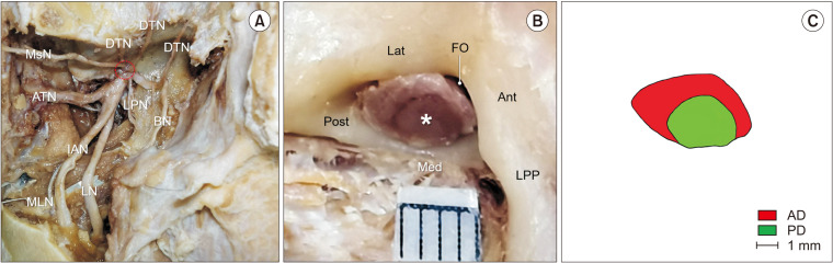
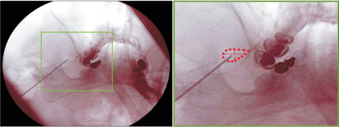
Background: Percutaneous radiofrequency thermocoagulation (RFTC) has been widely utilized in the management of trigeminal neuralgia. Despite using image guidance, accurate needle positioning into the target area still remains a critical element for achieving a successful outcome. This study was performed to precisely clarify the anatomical information required to ensure that the electrode tip is placed on the sensory component of the mandibular nerve (MN) at the foramen ovale (FO) level.
Methods: The study used 50 hemi-half heads from 26 South Korean adult cadavers.
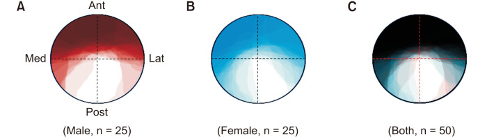
Results: The cross-sectioned anterior and posterior divisions of the MN at the FO level could be distinguished based on an irregular boundary and color difference. The anterior division was clearly brighter than the posterior one. The anterior division of the MN at the FO level was located at the whole anterior (38.0%), anteromedial (6.0%), anterior center (8.0%), and anterolateral (22.0%) parts. The posterior division was often located at the whole posterior or posterolateral parts of the MN at the FO level. The anterior divisions covered the whole MN except for the medial half of the posterolateral part in the overwrapped images of the cross-sectional areas of the MN at the FO level. The cross-sectional areas of the anterior divisions were similar in males and females, whereas those of the posterior divisions were significantly larger in males (P = 0.004).
Conclusions: The obtained anatomical information is expected to help physicians reduce unwanted side effects after percutaneous RFTC within the FO for the MN.
期刊介绍:
Korean Journal of Pain (Korean J Pain, KJP) is the official journal of the Korean Pain Society, founded in 1986. It has been published since 1988. It publishes peer reviewed original articles related to all aspects of pain, including clinical and basic research, patient care, education, and health policy. It has been published quarterly in English since 2009 (on the first day of January, April, July, and October). In addition, it has also become the official journal of the International Spinal Pain Society since 2016. The mission of the Journal is to improve the care of patients in pain by providing a forum for clinical researchers, basic scientists, clinicians, and other health professionals. The circulation number per issue is 50.
 求助内容:
求助内容: 应助结果提醒方式:


