{"title":"Prediction of Esophageal Varices in Viral Hepatitis C Cirrhosis: Performance of Combined Ultrasonography and Clinical Predictors.","authors":"Puwitch Charoenchue, Wittanee Na Chiangmai, Amonlaya Amantakul, Wasuwit Wanchaitanawong, Taned Chitapanarux, Suwalee Pojchamarnwiputh","doi":"10.1155/2023/7938732","DOIUrl":null,"url":null,"abstract":"<p><strong>Objectives: </strong>This study is aimed at evaluating the diagnostic performance of clinical predictors and the Doppler ultrasonography in predicting esophageal varices (EV) in patients with hepatitis C-related cirrhosis and exploring the practical predictors of EV.</p><p><strong>Methods: </strong>We conducted a prospective study from July 2020 to January 2021, enrolling 65 patients with mild hepatitis C-related cirrhosis. We obtained clinical data and performed grayscale and the Doppler ultrasound to explore the predictors of EV. Esophagogastroduodenoscopy (EGD) was performed as the reference test by the gastroenterologist within a week.</p><p><strong>Results: </strong>The prevalence of EV in the study was 41.5%. Multivariable regression analysis revealed that gender (female, OR = 4.04, <i>p</i> = 0.02), platelet count (<150000 per ml, OR = 3.13, <i>p</i> = 0.09), splenic length (>11 cm, OR = 3.64, <i>p</i> = 0.02), and absent right hepatic vein (RHV) triphasicity (OR = 3.15, <i>p</i> = 0.03) were significant predictors of EV. However, the diagnostic accuracy indices for isolated predictors were not good (AUROC = 0.63-0.66). A combination of these four predictors increases the diagnostic accuracy in predicting the presence of EV (AUROC = 0.80, 95% CI 0.69-0.91). Furthermore, the Doppler assessment of the right hepatic vein waveform showed good reproducibility (<i>κ</i> = 0.76).</p><p><strong>Conclusion: </strong>Combining clinical and Doppler ultrasound features can be used as a screening test for predicting the presence of EV in patients with hepatitis C-related cirrhosis. The practical predictors identified in this study could serve as an alternative to invasive EGD in EV diagnosis. Further studies are needed to explore the diagnostic accuracy of additional noninvasive predictors, such as elastography, to improve EV screening.</p>","PeriodicalId":47063,"journal":{"name":"International Journal of Biomedical Imaging","volume":"2023 ","pages":"7938732"},"PeriodicalIF":3.3000,"publicationDate":"2023-09-15","publicationTypes":"Journal Article","fieldsOfStudy":null,"isOpenAccess":false,"openAccessPdf":"https://www.ncbi.nlm.nih.gov/pmc/articles/PMC10516699/pdf/","citationCount":"0","resultStr":null,"platform":"Semanticscholar","paperid":null,"PeriodicalName":"International Journal of Biomedical Imaging","FirstCategoryId":"1085","ListUrlMain":"https://doi.org/10.1155/2023/7938732","RegionNum":0,"RegionCategory":null,"ArticlePicture":[],"TitleCN":null,"AbstractTextCN":null,"PMCID":null,"EPubDate":"2023/1/1 0:00:00","PubModel":"eCollection","JCR":"Q2","JCRName":"ENGINEERING, BIOMEDICAL","Score":null,"Total":0}
引用次数: 0
Abstract
Objectives: This study is aimed at evaluating the diagnostic performance of clinical predictors and the Doppler ultrasonography in predicting esophageal varices (EV) in patients with hepatitis C-related cirrhosis and exploring the practical predictors of EV.
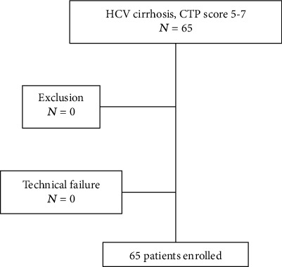
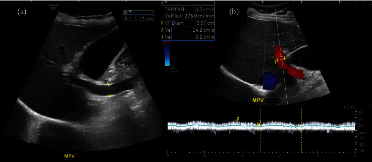
Methods: We conducted a prospective study from July 2020 to January 2021, enrolling 65 patients with mild hepatitis C-related cirrhosis. We obtained clinical data and performed grayscale and the Doppler ultrasound to explore the predictors of EV. Esophagogastroduodenoscopy (EGD) was performed as the reference test by the gastroenterologist within a week.
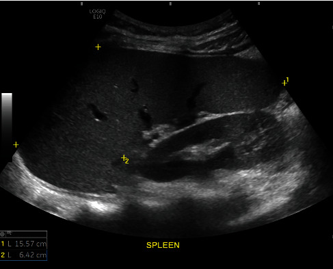
Results: The prevalence of EV in the study was 41.5%. Multivariable regression analysis revealed that gender (female, OR = 4.04, p = 0.02), platelet count (<150000 per ml, OR = 3.13, p = 0.09), splenic length (>11 cm, OR = 3.64, p = 0.02), and absent right hepatic vein (RHV) triphasicity (OR = 3.15, p = 0.03) were significant predictors of EV. However, the diagnostic accuracy indices for isolated predictors were not good (AUROC = 0.63-0.66). A combination of these four predictors increases the diagnostic accuracy in predicting the presence of EV (AUROC = 0.80, 95% CI 0.69-0.91). Furthermore, the Doppler assessment of the right hepatic vein waveform showed good reproducibility (κ = 0.76).
Conclusion: Combining clinical and Doppler ultrasound features can be used as a screening test for predicting the presence of EV in patients with hepatitis C-related cirrhosis. The practical predictors identified in this study could serve as an alternative to invasive EGD in EV diagnosis. Further studies are needed to explore the diagnostic accuracy of additional noninvasive predictors, such as elastography, to improve EV screening.
期刊介绍:
The International Journal of Biomedical Imaging is managed by a board of editors comprising internationally renowned active researchers. The journal is freely accessible online and also offered for purchase in print format. It employs a web-based review system to ensure swift turnaround times while maintaining high standards. In addition to regular issues, special issues are organized by guest editors. The subject areas covered include (but are not limited to):
Digital radiography and tomosynthesis
X-ray computed tomography (CT)
Magnetic resonance imaging (MRI)
Single photon emission computed tomography (SPECT)
Positron emission tomography (PET)
Ultrasound imaging
Diffuse optical tomography, coherence, fluorescence, bioluminescence tomography, impedance tomography
Neutron imaging for biomedical applications
Magnetic and optical spectroscopy, and optical biopsy
Optical, electron, scanning tunneling/atomic force microscopy
Small animal imaging
Functional, cellular, and molecular imaging
Imaging assays for screening and molecular analysis
Microarray image analysis and bioinformatics
Emerging biomedical imaging techniques
Imaging modality fusion
Biomedical imaging instrumentation
Biomedical image processing, pattern recognition, and analysis
Biomedical image visualization, compression, transmission, and storage
Imaging and modeling related to systems biology and systems biomedicine
Applied mathematics, applied physics, and chemistry related to biomedical imaging
Grid-enabling technology for biomedical imaging and informatics

 求助内容:
求助内容: 应助结果提醒方式:


