Hualin Xu, Jie Fu, Qiang Tu, Qingyun Shuai, Yizhi Chen, Fuyun Wu, Zheng Cao
{"title":"The SGLT2 inhibitor empagliflozin attenuates atherosclerosis progression by inducing autophagy.","authors":"Hualin Xu, Jie Fu, Qiang Tu, Qingyun Shuai, Yizhi Chen, Fuyun Wu, Zheng Cao","doi":"10.1007/s13105-023-00974-0","DOIUrl":null,"url":null,"abstract":"<p><p>Cardiovascular disease due to atherosclerosis is one of the leading causes of death worldwide; however, the underlying mechanism has yet to be defined. The sodium-dependent glucose transporter 2 inhibitor (SGLT2i) empagliflozin is a new type of hypoglycemic drug. Recent studies have shown that empagliflozin not only reduces high glucose levels but also exerts cardiovascular-protective effects and slows the process of atherosclerosis. The purpose of this study was to elucidate the mechanism by which empagliflozin ameliorates atherosclerosis. Male apolipoprotein E-deficient (ApoE<sup>-/-</sup>) mice were fed a high-fat Western diet to establish an atherosclerosis model. The area and size of atherosclerotic lesions in ApoE<sup>-/-</sup> mice were then assessed by performing hematoxylin-eosin (HE) staining after empagliflozin treatment. Concurrently, oxidized low-density lipoprotein (oxLDL) was used to mimic atherosclerosis in three different types of cells. Then, following empagliflozin treatment of macrophage cells (RAW264.7), human aortic smooth muscle cells (HASMCs), and human umbilical vein endothelial cells (HUVECs), western blotting was applied to measure the levels of autophagy-related proteins and proinflammatory cytokines, and green fluorescent protein (GFP)-light chain 3 (LC3) puncta were detected using confocal microscopy to confirm autophagosome formation. Oil Red O staining was performed to detect the foaming of macrophages and HASMCs, and flow cytometry was used for the cell cycle analysis. 5-ethynyl-2'-deoxyuridine (EdU), cell counting kit-8 (CCK-8), and scratch assays were also performed to examine the proliferation and migration of HASMCs. Empagliflozin suppressed the progression of atherosclerotic lesions in ApoE<sup>-/-</sup> mice. Empagliflozin also induced autophagy in RAW246.7 cells, HASMCs, and HUVECs via the adenosine monophosphate-activated protein kinase (AMPK) signaling pathway, and it significantly increased the levels of the Beclin1 protein, the LC3B-II/I ratio, and p-AMPK protein. In addition, empagliflozin decreased the expression of P62 and the protein levels of inflammatory cytokines, and it inhibited the foaming of RAW246.7 cells and HASMCs, as well as the expression of inflammatory factors by inducing autophagy. Empagliflozin activated autophagy through the AMPK signaling pathway to delay the progression of atherosclerosis. Furthermore, the results of flow cytometry, EdU assays, CCK-8 cell viability assays, and scratch assays indicated that empagliflozin blocked HASMCs proliferation and migration. Empagliflozin activates autophagy through the AMPK signaling pathway to delay the evolution of atherosclerosis, indicating that it may represent a new and effective drug for the clinical treatment of atherosclerosis.</p>","PeriodicalId":16779,"journal":{"name":"Journal of physiology and biochemistry","volume":" ","pages":"27-39"},"PeriodicalIF":3.7000,"publicationDate":"2024-02-01","publicationTypes":"Journal Article","fieldsOfStudy":null,"isOpenAccess":false,"openAccessPdf":"","citationCount":"0","resultStr":null,"platform":"Semanticscholar","paperid":null,"PeriodicalName":"Journal of physiology and biochemistry","FirstCategoryId":"99","ListUrlMain":"https://doi.org/10.1007/s13105-023-00974-0","RegionNum":3,"RegionCategory":"生物学","ArticlePicture":[],"TitleCN":null,"AbstractTextCN":null,"PMCID":null,"EPubDate":"2023/10/4 0:00:00","PubModel":"Epub","JCR":"Q2","JCRName":"BIOCHEMISTRY & MOLECULAR BIOLOGY","Score":null,"Total":0}
引用次数: 0
Abstract
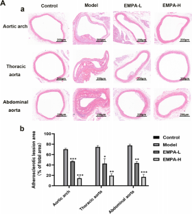
Cardiovascular disease due to atherosclerosis is one of the leading causes of death worldwide; however, the underlying mechanism has yet to be defined. The sodium-dependent glucose transporter 2 inhibitor (SGLT2i) empagliflozin is a new type of hypoglycemic drug. Recent studies have shown that empagliflozin not only reduces high glucose levels but also exerts cardiovascular-protective effects and slows the process of atherosclerosis. The purpose of this study was to elucidate the mechanism by which empagliflozin ameliorates atherosclerosis. Male apolipoprotein E-deficient (ApoE-/-) mice were fed a high-fat Western diet to establish an atherosclerosis model. The area and size of atherosclerotic lesions in ApoE-/- mice were then assessed by performing hematoxylin-eosin (HE) staining after empagliflozin treatment. Concurrently, oxidized low-density lipoprotein (oxLDL) was used to mimic atherosclerosis in three different types of cells. Then, following empagliflozin treatment of macrophage cells (RAW264.7), human aortic smooth muscle cells (HASMCs), and human umbilical vein endothelial cells (HUVECs), western blotting was applied to measure the levels of autophagy-related proteins and proinflammatory cytokines, and green fluorescent protein (GFP)-light chain 3 (LC3) puncta were detected using confocal microscopy to confirm autophagosome formation. Oil Red O staining was performed to detect the foaming of macrophages and HASMCs, and flow cytometry was used for the cell cycle analysis. 5-ethynyl-2'-deoxyuridine (EdU), cell counting kit-8 (CCK-8), and scratch assays were also performed to examine the proliferation and migration of HASMCs. Empagliflozin suppressed the progression of atherosclerotic lesions in ApoE-/- mice. Empagliflozin also induced autophagy in RAW246.7 cells, HASMCs, and HUVECs via the adenosine monophosphate-activated protein kinase (AMPK) signaling pathway, and it significantly increased the levels of the Beclin1 protein, the LC3B-II/I ratio, and p-AMPK protein. In addition, empagliflozin decreased the expression of P62 and the protein levels of inflammatory cytokines, and it inhibited the foaming of RAW246.7 cells and HASMCs, as well as the expression of inflammatory factors by inducing autophagy. Empagliflozin activated autophagy through the AMPK signaling pathway to delay the progression of atherosclerosis. Furthermore, the results of flow cytometry, EdU assays, CCK-8 cell viability assays, and scratch assays indicated that empagliflozin blocked HASMCs proliferation and migration. Empagliflozin activates autophagy through the AMPK signaling pathway to delay the evolution of atherosclerosis, indicating that it may represent a new and effective drug for the clinical treatment of atherosclerosis.
期刊介绍:
The Journal of Physiology and Biochemistry publishes original research articles and reviews describing relevant new observations on molecular, biochemical and cellular mechanisms involved in human physiology. All areas of the physiology are covered. Special emphasis is placed on the integration of those levels in the whole-organism. The Journal of Physiology and Biochemistry also welcomes articles on molecular nutrition and metabolism studies, and works related to the genomic or proteomic bases of the physiological functions. Descriptive manuscripts about physiological/biochemical processes or clinical manuscripts will not be considered. The journal will not accept manuscripts testing effects of animal or plant extracts.
 求助内容:
求助内容: 应助结果提醒方式:


