{"title":"Changed cortical thickness and sulcal depth in pediatric acute lymphoblastic leukemia survivors treated with chemotherapy only.","authors":"Fangling Li, Yimin Guo, Gaoqiang Xu, Ying Liu, Xiaoxi Chen, Tijiang Zhang","doi":"10.1007/s11682-023-00794-2","DOIUrl":null,"url":null,"abstract":"<p><p>The purpose of this study is to observe the changes of cortical morphological characteristics and their potential contribution to cognitive function in ALL survivors by using surface-based morphometry (SBM). Using SBM analysis, we calculated and compared group differences in cortical thickness, sulcal depth, gyrification, and fractal dimension of the cerebral cortex between 18 pediatric ALL survivors treated on chemotherapy-only protocols and off treatment within 2 years, and 18 healthy controls (HCs) with two-sample t-tests [P < 0.05, family-wise error (FWE) corrected]. Relationships between abnormal cortical characteristic values and cognitive function parameters were investigated with partial correlation analysis, taking age as a covariate. We found decreased cortical thickness mainly located in the prefrontal and temporal region, and increased sulcal depth in left rostral middle frontal cortex and left pars orbitalis in the ALL survivors compared to HCs. There were no statistically significant differences in the gyrification and fractal dimension between the two groups. In ALL survivors, cortical thickness and sulcal depth of above areas values revealed no significant correlation with the cognitive function parameters. In conclusion, pediatric ALL survivors show decreased cortical thickness in prefrontal and temporal regions, and increased sulcal depth in prefrontal region. These results suggest that SBM-based approach can be used to assess changes of cortical morphological characteristics in pediatric ALL survivors.</p>","PeriodicalId":9192,"journal":{"name":"Brain Imaging and Behavior","volume":" ","pages":"738-748"},"PeriodicalIF":2.4000,"publicationDate":"2023-12-01","publicationTypes":"Journal Article","fieldsOfStudy":null,"isOpenAccess":false,"openAccessPdf":"","citationCount":"0","resultStr":null,"platform":"Semanticscholar","paperid":null,"PeriodicalName":"Brain Imaging and Behavior","FirstCategoryId":"3","ListUrlMain":"https://doi.org/10.1007/s11682-023-00794-2","RegionNum":3,"RegionCategory":"医学","ArticlePicture":[],"TitleCN":null,"AbstractTextCN":null,"PMCID":null,"EPubDate":"2023/9/22 0:00:00","PubModel":"Epub","JCR":"Q2","JCRName":"NEUROIMAGING","Score":null,"Total":0}
引用次数: 0
Abstract
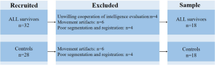
The purpose of this study is to observe the changes of cortical morphological characteristics and their potential contribution to cognitive function in ALL survivors by using surface-based morphometry (SBM). Using SBM analysis, we calculated and compared group differences in cortical thickness, sulcal depth, gyrification, and fractal dimension of the cerebral cortex between 18 pediatric ALL survivors treated on chemotherapy-only protocols and off treatment within 2 years, and 18 healthy controls (HCs) with two-sample t-tests [P < 0.05, family-wise error (FWE) corrected]. Relationships between abnormal cortical characteristic values and cognitive function parameters were investigated with partial correlation analysis, taking age as a covariate. We found decreased cortical thickness mainly located in the prefrontal and temporal region, and increased sulcal depth in left rostral middle frontal cortex and left pars orbitalis in the ALL survivors compared to HCs. There were no statistically significant differences in the gyrification and fractal dimension between the two groups. In ALL survivors, cortical thickness and sulcal depth of above areas values revealed no significant correlation with the cognitive function parameters. In conclusion, pediatric ALL survivors show decreased cortical thickness in prefrontal and temporal regions, and increased sulcal depth in prefrontal region. These results suggest that SBM-based approach can be used to assess changes of cortical morphological characteristics in pediatric ALL survivors.
期刊介绍:
Brain Imaging and Behavior is a bi-monthly, peer-reviewed journal, that publishes clinically relevant research using neuroimaging approaches to enhance our understanding of disorders of higher brain function. The journal is targeted at clinicians and researchers in fields concerned with human brain-behavior relationships, such as neuropsychology, psychiatry, neurology, neurosurgery, rehabilitation, and cognitive neuroscience.

 求助内容:
求助内容: 应助结果提醒方式:


