Gamze Ucan Gunduz, Aysegul Mavi Yildiz, Ozgur Yalcinbayir, Mehmet Baykara, Esin Sogutlu Sari, Sevde Isleker, Nilufer Aylin Acet Ozturk
{"title":"Pupillographic Analysis of COVID-19 Patients: Early and Late Results After Recovery.","authors":"Gamze Ucan Gunduz, Aysegul Mavi Yildiz, Ozgur Yalcinbayir, Mehmet Baykara, Esin Sogutlu Sari, Sevde Isleker, Nilufer Aylin Acet Ozturk","doi":"10.14744/bej.2023.30592","DOIUrl":null,"url":null,"abstract":"<p><strong>Objectives: </strong>We aimed to investigate the short- and long-term static and dynamic pupillary responses of patients recovered from coronavirus disease-19 (COVID-19) using quantitative infrared pupillography.</p><p><strong>Methods: </strong>This study included patients who recovered from COVID-19 (Group 1) and age- and gender-matched controls (Group 2). A detailed ophthalmic examination was performed at 1 month and 6 months after the diagnosis of COVID-19. Photopic, mesopic, and scotopic pupil diameters (PDs) were measured using a quantitative infrared pupillography which was integrated into Scheimpflug/Placido photography-based topography system. PDs at 0, 2<sup>nd</sup>, 4<sup>th</sup>, and 6<sup>th</sup> seconds, and average pupil dilation speeds at 2<sup>nd</sup>, 4<sup>th</sup>, 6<sup>th</sup>, and 8<sup>th</sup> seconds were recorded.</p><p><strong>Results: </strong>Eighty-six eyes of 86 patients (Group 1: n=42; Group 2: n=44) were included. While the mean photopic, mesopic, and scotopic PDs were significantly larger in the COVID-19 group than the control group in the 1<sup>st</sup> month (p=0.035, p=0.017, p=0.018, respectively), no statistically significant difference was found in the 6<sup>th</sup> month. Besides, average pupil dilation speeds and PDs at the 0, 2<sup>nd</sup>, 4<sup>th</sup>, and 6<sup>th</sup> seconds were not statistically significantly different between the two groups in the 1<sup>st</sup> month and 6<sup>th</sup> month.</p><p><strong>Conclusion: </strong>PDs were significantly larger in COVID-19 patients in all light intensities in the 1<sup>st</sup> month after COVID-19. However, pupillary dilation was transient, and no significant difference was found in the 6<sup>th</sup> month. We suggest that the transient pupillary dilation may be secondary to the autonomic nervous system dysfunction and/or optic nerve and visual pathways alterations following COVID-19.</p>","PeriodicalId":8740,"journal":{"name":"Beyoglu Eye Journal","volume":"8 3","pages":"149-156"},"PeriodicalIF":0.0000,"publicationDate":"2023-09-13","publicationTypes":"Journal Article","fieldsOfStudy":null,"isOpenAccess":false,"openAccessPdf":"https://ftp.ncbi.nlm.nih.gov/pub/pmc/oa_pdf/27/a5/BEJ-8-149.PMC10521134.pdf","citationCount":"0","resultStr":null,"platform":"Semanticscholar","paperid":null,"PeriodicalName":"Beyoglu Eye Journal","FirstCategoryId":"1085","ListUrlMain":"https://doi.org/10.14744/bej.2023.30592","RegionNum":0,"RegionCategory":null,"ArticlePicture":[],"TitleCN":null,"AbstractTextCN":null,"PMCID":null,"EPubDate":"2023/1/1 0:00:00","PubModel":"eCollection","JCR":"","JCRName":"","Score":null,"Total":0}
引用次数: 0
Abstract
Objectives: We aimed to investigate the short- and long-term static and dynamic pupillary responses of patients recovered from coronavirus disease-19 (COVID-19) using quantitative infrared pupillography.
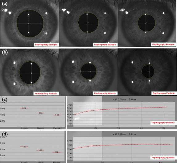
Methods: This study included patients who recovered from COVID-19 (Group 1) and age- and gender-matched controls (Group 2). A detailed ophthalmic examination was performed at 1 month and 6 months after the diagnosis of COVID-19. Photopic, mesopic, and scotopic pupil diameters (PDs) were measured using a quantitative infrared pupillography which was integrated into Scheimpflug/Placido photography-based topography system. PDs at 0, 2nd, 4th, and 6th seconds, and average pupil dilation speeds at 2nd, 4th, 6th, and 8th seconds were recorded.
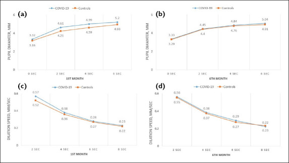
Results: Eighty-six eyes of 86 patients (Group 1: n=42; Group 2: n=44) were included. While the mean photopic, mesopic, and scotopic PDs were significantly larger in the COVID-19 group than the control group in the 1st month (p=0.035, p=0.017, p=0.018, respectively), no statistically significant difference was found in the 6th month. Besides, average pupil dilation speeds and PDs at the 0, 2nd, 4th, and 6th seconds were not statistically significantly different between the two groups in the 1st month and 6th month.
Conclusion: PDs were significantly larger in COVID-19 patients in all light intensities in the 1st month after COVID-19. However, pupillary dilation was transient, and no significant difference was found in the 6th month. We suggest that the transient pupillary dilation may be secondary to the autonomic nervous system dysfunction and/or optic nerve and visual pathways alterations following COVID-19.

 求助内容:
求助内容: 应助结果提醒方式:


