{"title":"Psoriasis Area and Severity Index (PASI) Objectivisation by Flow Cytometry Analysis of Major Lymphocytes Subsets.","authors":"Lamija Zecevic-Pasic, Nejra Dzananovic, Refet Gojak, Suzana Tihic-Kapidzic, Berina Hasanefendic, Ermin Begovic, Amir Fazlagic","doi":"10.5455/aim.2023.31.206-210","DOIUrl":null,"url":null,"abstract":"<p><strong>Background: </strong>Psoriasis as an immune-mediated inflammatory skin disease. The basis of the pathogenesis of psoriasis is the dysregulation of immune cell function in genetically predisposed individuals. The characteristic dysfunction of the immune system in patients with psoriasis is manifested as a variation in the cellular phenotypic profile in accordance with the disease status.</p><p><strong>Objective: </strong>The aim of this study was to evaluate the immunophenotypic profile of lymphocytes obtained by flow cytometry as an auxiliary diagnostic tool in the objectivization of the PASI score.</p><p><strong>Methods: </strong>The study group included 40 patients with psoriasis, hospitalized and treated at Dermatology Clinic of Clinical center University of Sarajevo and 30 healthy individuals as controls. After venepunction, the blood samples for determining the immune profile were prepared following standard laboratory procedures using conjugated monoclonal antibodies and BD FACSCanto II flow cytometer. T-lymphocytes (CD3, CD4, CD8), B lymphocytes (CD19), Natural killer cells (NK), and activatet T-cells (CD3HLA) were determined for all patients. Based on the PASI score, the severity and area of the disease was assessed for all psoriasis patients by dermatology specialist.</p><p><strong>Results: </strong>Our data shows no significant difference in any of the lymphocyte subpopulations between psoriasis patients and healthy controls, except CD3HLA. CD3HLA has higher values in patients with psoriasis, p=0.015. Of all the parameters, only NK cells were significantly correlated to the PASI score (rho -0.279; p=0.048). ROC curve analysis revealed a statistically significant difference for the proportion of CD3 lymphocytes (AUC 0.799; p=0.004), CD8 lymphocytes (AUC 0.733; p= 0.023), NK cells (AUC 0.722; p=0.008) and CD3HLA activated T lymphocytes (AUC 0.347; p=0.034).</p><p><strong>Conclusion: </strong>Profile of major lymphocyte subsets in patients with psoriasis is similar to that of healthy controls. The values of CD3, CD8, NK, CD3HLA were defined as biomarkers capable of distinguishing psoriasis according to the severity of the disease. Immunophenotyping of peripheral blood lymphocytes can play an important role as an auxiliary diagnostic method in differentiating the clinical stages of psoriasis and objectifying the PASI score.</p>","PeriodicalId":7074,"journal":{"name":"Acta Informatica Medica","volume":"31 3","pages":"206-210"},"PeriodicalIF":0.0000,"publicationDate":"2023-01-01","publicationTypes":"Journal Article","fieldsOfStudy":null,"isOpenAccess":false,"openAccessPdf":"https://ftp.ncbi.nlm.nih.gov/pub/pmc/oa_pdf/85/2f/AIM-31-206.PMC10540746.pdf","citationCount":"0","resultStr":null,"platform":"Semanticscholar","paperid":null,"PeriodicalName":"Acta Informatica Medica","FirstCategoryId":"1085","ListUrlMain":"https://doi.org/10.5455/aim.2023.31.206-210","RegionNum":0,"RegionCategory":null,"ArticlePicture":[],"TitleCN":null,"AbstractTextCN":null,"PMCID":null,"EPubDate":"","PubModel":"","JCR":"Q2","JCRName":"Medicine","Score":null,"Total":0}
引用次数: 0
Abstract
Background: Psoriasis as an immune-mediated inflammatory skin disease. The basis of the pathogenesis of psoriasis is the dysregulation of immune cell function in genetically predisposed individuals. The characteristic dysfunction of the immune system in patients with psoriasis is manifested as a variation in the cellular phenotypic profile in accordance with the disease status.
Objective: The aim of this study was to evaluate the immunophenotypic profile of lymphocytes obtained by flow cytometry as an auxiliary diagnostic tool in the objectivization of the PASI score.
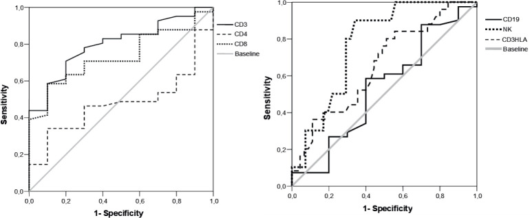
Methods: The study group included 40 patients with psoriasis, hospitalized and treated at Dermatology Clinic of Clinical center University of Sarajevo and 30 healthy individuals as controls. After venepunction, the blood samples for determining the immune profile were prepared following standard laboratory procedures using conjugated monoclonal antibodies and BD FACSCanto II flow cytometer. T-lymphocytes (CD3, CD4, CD8), B lymphocytes (CD19), Natural killer cells (NK), and activatet T-cells (CD3HLA) were determined for all patients. Based on the PASI score, the severity and area of the disease was assessed for all psoriasis patients by dermatology specialist.
Results: Our data shows no significant difference in any of the lymphocyte subpopulations between psoriasis patients and healthy controls, except CD3HLA. CD3HLA has higher values in patients with psoriasis, p=0.015. Of all the parameters, only NK cells were significantly correlated to the PASI score (rho -0.279; p=0.048). ROC curve analysis revealed a statistically significant difference for the proportion of CD3 lymphocytes (AUC 0.799; p=0.004), CD8 lymphocytes (AUC 0.733; p= 0.023), NK cells (AUC 0.722; p=0.008) and CD3HLA activated T lymphocytes (AUC 0.347; p=0.034).
Conclusion: Profile of major lymphocyte subsets in patients with psoriasis is similar to that of healthy controls. The values of CD3, CD8, NK, CD3HLA were defined as biomarkers capable of distinguishing psoriasis according to the severity of the disease. Immunophenotyping of peripheral blood lymphocytes can play an important role as an auxiliary diagnostic method in differentiating the clinical stages of psoriasis and objectifying the PASI score.

 求助内容:
求助内容: 应助结果提醒方式:


