Age-related changes in muscle thickness, echo intensity and shear modulus of the iliocapsularis
Abstract
Purpose
This study aimed to clarify age-related changes in the iliocapsularis (IC) using indicators of quantity, quality, and mechanical properties. We also compared the age-related changes in the IC and other hip muscles.
Methods
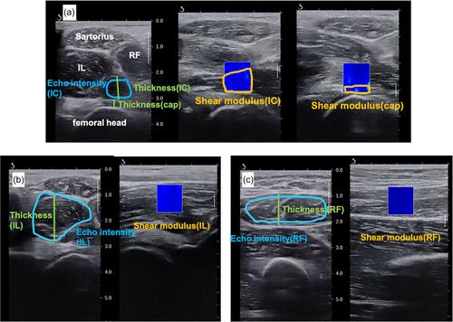
Eighty-seven healthy women (ages: 21–82 years, mean age: 45.9 ± 15.7 years) participated in the experiment. We measured thickness, echo intensity, and shear modulus of the IC, iliacus muscle, rectus femoris, and the thickness and shear modulus of the hip joint capsule. Spearman's rank correlation coefficient was used to measure the association of age with variables measured in the muscles and joint capsule.
Results
Thickness of the iliacus muscle and rectus femoris decreased significantly with age, but the thickness of the IC and hip joint capsule showed no significant correlation. The echo intensities of the IC, iliacus muscle, and rectus femoris were positively correlated, which increased with age. Furthermore, the shear modulus of the iliacus, rectus femoris, and hip joint capsule showed an increase with age, whereas the shear modulus of the IC exhibited no correlation with age.
Conclusion
The muscle quality of the IC changed significantly, unlike that of the iliacus or rectus femoris. Additionally, the correlation with echo intensity was relatively weaker in the IC compared with the iliacus or rectus femoris.
 求助内容:
求助内容: 应助结果提醒方式:


