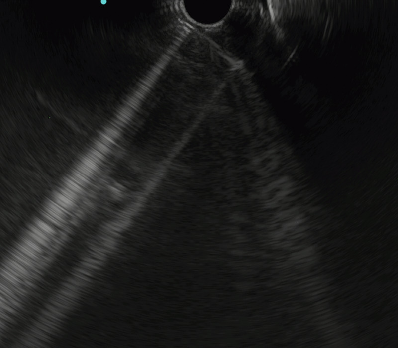
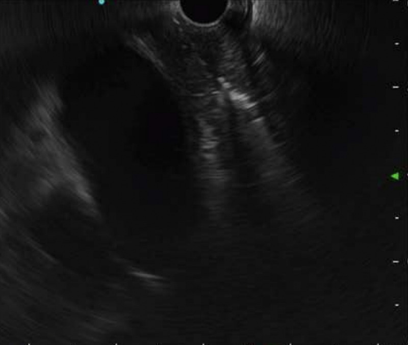
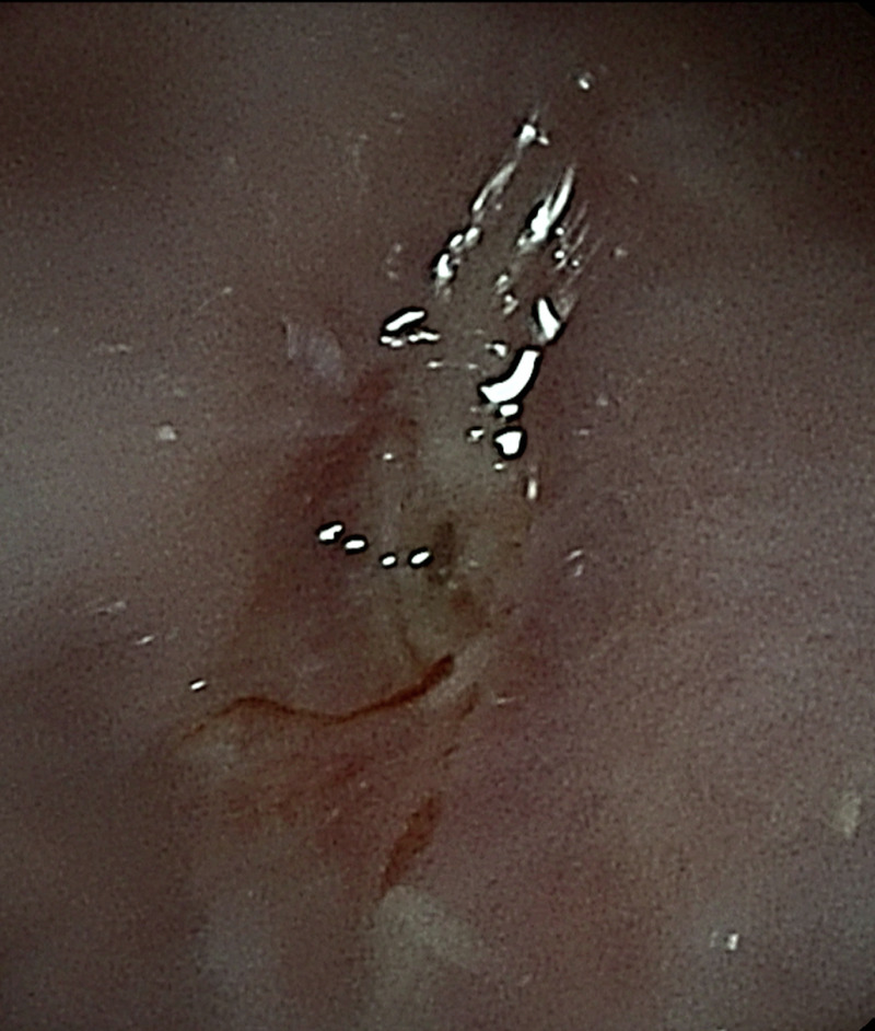
EUS-guided esophageal lumen restoration in a young patient with complete luminal obstruction (with video).
IF 5.4
1区 医学
Q1 GASTROENTEROLOGY & HEPATOLOGY
引用次数: 0
Abstract
A 28-year-old woman with type 1 diabetes presented with severe dysphagia after pregnancy. Endoscopy showed a “ blind ” esophagus, with complete esophageal obstruction [Figure 1]. An endoscopic approach was unsuccessful in finding an access for the guidewire, and no evidence of contrast medium passage was seen. EUS was used to study the esophageal layers and try to see behind the stricture. The wall was thickened (10 mm), with prevalence of the submucosa. A mild insufflation of air, used as ultrasonographic contrast, allowed the endosonographer to see a hyperechoic line that was interpreted as the submillimetric remnant of the esophageal lumen [Figure 2]. A 19-gauge access needle was used to puncture, under EUS guidance, the esophageal wall [Figure 3] starting from the hyperechoic line inside the esophagus. After initial resistance, under fluoroscopic view of the needle path, a point of least resistance was felt, and the needle tip, inserted for almost 5 cm, was visualized below the diaphragm. A guidewire was inserted into the needle and under fluoroscopic view was seen creating a loop inside the stomach [Figure 4]. A Hurricane RX biliary balloon dilation catheter (6-mm diameter, 4-cm length), (Boston Scientific Corp., Natick, MA) was passed through the stricture, and the first dilation was performed [Figure 5]. In the following weeks, other dilations were performed with Savary bougies (Boston Scientific Corp., Natick, MA), and the esophageal lumen was restored [Video 1]. Forceps biopsies revealed acute and chronic inflammation, with severe fibrosis and no signs of


EUS引导下一名完全性食管管腔梗阻的年轻患者进行食管管腔修复(附视频)。
本文章由计算机程序翻译,如有差异,请以英文原文为准。
求助全文
约1分钟内获得全文
求助全文
来源期刊

Endoscopic Ultrasound
GASTROENTEROLOGY & HEPATOLOGY-
CiteScore
6.20
自引率
11.10%
发文量
144
期刊介绍:
Endoscopic Ultrasound, a publication of Euro-EUS Scientific Committee, Asia-Pacific EUS Task Force and Latin American Chapter of EUS, is a peer-reviewed online journal with Quarterly print on demand compilation of issues published. The journal’s full text is available online at http://www.eusjournal.com. The journal allows free access (Open Access) to its contents and permits authors to self-archive final accepted version of the articles on any OAI-compliant institutional / subject-based repository. The journal does not charge for submission, processing or publication of manuscripts and even for color reproduction of photographs.
 求助内容:
求助内容: 应助结果提醒方式:


