The prophylactic value of TNF-α inhibitors against retinal cell apoptosis and optic nerve axon loss after corneal surgery or trauma

IF 3 3区 医学 Q1 OPHTHALMOLOGY
Eleftherios I. Paschalis, Chengxin Zhou, Jyoti Sharma, Thomas H. Dohlman, Sarah Kim, Fengyang Lei, James Chodosh, Demetrios Vavvas, Arto Urtti, George Papaliodis, Claes H. Dohlman
{"title":"The prophylactic value of TNF-α inhibitors against retinal cell apoptosis and optic nerve axon loss after corneal surgery or trauma","authors":"Eleftherios I. Paschalis,&nbsp;Chengxin Zhou,&nbsp;Jyoti Sharma,&nbsp;Thomas H. Dohlman,&nbsp;Sarah Kim,&nbsp;Fengyang Lei,&nbsp;James Chodosh,&nbsp;Demetrios Vavvas,&nbsp;Arto Urtti,&nbsp;George Papaliodis,&nbsp;Claes H. Dohlman","doi":"10.1111/aos.15786","DOIUrl":null,"url":null,"abstract":"<div>\n \n \n <section>\n \n <h3> Background and Purpose</h3>\n \n <p>Late secondary glaucoma is an often-severe complication after acute events like anterior segment surgery, trauma and infection. TNF-α is a major mediator that is rapidly upregulated, diffusing also to the retina and causes apoptosis of the ganglion cells and degeneration of their optic nerve axons (mediating steps to glaucomatous damage). Anti-TNF-α antibodies are in animals very effective in protecting the retinal cells and the optic nerve—and might therefore be useful prophylactically against secondary glaucoma in future such patients. Here we evaluate (1) <i>toxicity</i> and (2) <i>efficacy</i> of two TNF-α inhibitors (adalimumab and infliximab), in rabbits by <i>subconjunctival</i> administration.</p>\n </section>\n \n <section>\n \n <h3> Methods</h3>\n \n <p>For drug <i>toxicity</i>, animals with <i>normal, unburned</i> corneas were injected with adalimumab (0.4, 4, or 40 mg), or infliximab (1, 10, or 100 mg). For drug <i>efficacy</i>, other animals were subjected to alkali burn before such injection, or steroids (for control). The rabbits were evaluated clinically with slit lamp and photography, electroretinography, optical coherence tomography, and intraocular pressure manometry. A sub-set of eyes were stained ex vivo after 3 days for retinal cell apoptosis (TUNEL). In other experiments the optic nerves were evaluated by paraphenylenediamine staining after 50 or 90 days. Loss of retinal cells and optic nerve degeneration were quantified.</p>\n </section>\n \n <section>\n \n <h3> Results</h3>\n \n <p>Subconjunctival administration of 0.4 mg or 4.0 mg adalimumab were well tolerated, whereas 40.0 mg was toxic to the retina. 1, 10, or 100 mg infliximab were also well tolerated. Analysis of the optic nerve axons after 50 days confirmed the safety of 4.0 mg adalimumab and of 100 mg infliximab. For <i>efficacy, 4.0 mg adalimumab subconjunctivally in 0.08 mL</i> provided practically full protection against retinal cell apoptosis 3 days following alkali burn, and infliximab 100 mg only slightly less. At 90 days following burn injury, control optic nerves showed about 50% axon loss as compared to 8% in the adalimumab treatment group.</p>\n </section>\n \n <section>\n \n <h3> Conclusions</h3>\n \n <p><i>Subconjunctival injection of 4.0 mg adalimumab</i> in rabbits shows no eye toxicity and provides excellent neuroprotection, both short (3 days) and long-term (90 days). Our total. accumulated data from several of our studies, combined with the present paper, suggest that corneal injuries, including surgery, might benefit from routine administration of anti-TNF-α biologics to reduce inflammation and future secondary glaucoma.</p>\n </section>\n </div>","PeriodicalId":6915,"journal":{"name":"Acta Ophthalmologica","volume":"102 3","pages":"e381-e394"},"PeriodicalIF":3.0000,"publicationDate":"2023-10-06","publicationTypes":"Journal Article","fieldsOfStudy":null,"isOpenAccess":false,"openAccessPdf":"https://onlinelibrary.wiley.com/doi/epdf/10.1111/aos.15786","citationCount":"0","resultStr":null,"platform":"Semanticscholar","paperid":null,"PeriodicalName":"Acta Ophthalmologica","FirstCategoryId":"3","ListUrlMain":"https://onlinelibrary.wiley.com/doi/10.1111/aos.15786","RegionNum":3,"RegionCategory":"医学","ArticlePicture":[],"TitleCN":null,"AbstractTextCN":null,"PMCID":null,"EPubDate":"","PubModel":"","JCR":"Q1","JCRName":"OPHTHALMOLOGY","Score":null,"Total":0}
引用次数: 0

Abstract

Background and Purpose

Late secondary glaucoma is an often-severe complication after acute events like anterior segment surgery, trauma and infection. TNF-α is a major mediator that is rapidly upregulated, diffusing also to the retina and causes apoptosis of the ganglion cells and degeneration of their optic nerve axons (mediating steps to glaucomatous damage). Anti-TNF-α antibodies are in animals very effective in protecting the retinal cells and the optic nerve—and might therefore be useful prophylactically against secondary glaucoma in future such patients. Here we evaluate (1) toxicity and (2) efficacy of two TNF-α inhibitors (adalimumab and infliximab), in rabbits by subconjunctival administration.

Methods

For drug toxicity, animals with normal, unburned corneas were injected with adalimumab (0.4, 4, or 40 mg), or infliximab (1, 10, or 100 mg). For drug efficacy, other animals were subjected to alkali burn before such injection, or steroids (for control). The rabbits were evaluated clinically with slit lamp and photography, electroretinography, optical coherence tomography, and intraocular pressure manometry. A sub-set of eyes were stained ex vivo after 3 days for retinal cell apoptosis (TUNEL). In other experiments the optic nerves were evaluated by paraphenylenediamine staining after 50 or 90 days. Loss of retinal cells and optic nerve degeneration were quantified.

Results

Subconjunctival administration of 0.4 mg or 4.0 mg adalimumab were well tolerated, whereas 40.0 mg was toxic to the retina. 1, 10, or 100 mg infliximab were also well tolerated. Analysis of the optic nerve axons after 50 days confirmed the safety of 4.0 mg adalimumab and of 100 mg infliximab. For efficacy, 4.0 mg adalimumab subconjunctivally in 0.08 mL provided practically full protection against retinal cell apoptosis 3 days following alkali burn, and infliximab 100 mg only slightly less. At 90 days following burn injury, control optic nerves showed about 50% axon loss as compared to 8% in the adalimumab treatment group.

Conclusions

Subconjunctival injection of 4.0 mg adalimumab in rabbits shows no eye toxicity and provides excellent neuroprotection, both short (3 days) and long-term (90 days). Our total. accumulated data from several of our studies, combined with the present paper, suggest that corneal injuries, including surgery, might benefit from routine administration of anti-TNF-α biologics to reduce inflammation and future secondary glaucoma.
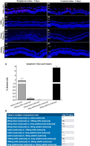
Abstract Image

TNF-α抑制剂对角膜手术或创伤后视网膜细胞凋亡和视神经轴突丢失的预防价值。
背景和目的:晚期继发性青光眼是眼前段手术、创伤和感染等急性事件后常见的严重并发症。TNF-α是一种快速上调的主要介质,也会扩散到视网膜,并导致神经节细胞凋亡和视神经轴突变性(介导青光眼损伤的步骤)。抗TNF-α抗体在动物体内对保护视网膜细胞和视神经非常有效,因此可能对未来此类患者的继发性青光眼有预防作用。在这里,我们通过结膜下给药评估了(1)两种TNF-α抑制剂(阿达木单抗和英夫利昔单抗)在兔体内的毒性和(2)疗效。方法:对于药物毒性,用阿达木单抗(0.4、4或40 mg),或英夫利昔单抗(1、10或100 mg)。对于药物疗效,其他动物在注射前进行碱烧伤,或使用类固醇(用于对照)。通过裂隙灯和摄影、视网膜电图、光学相干断层扫描和眼压计对兔子进行临床评估。一组眼睛在3天后进行离体染色 天进行视网膜细胞凋亡(TUNEL)。在其他实验中,在50或90后通过对苯二胺染色来评估视神经 天。对视网膜细胞损失和视神经变性进行定量。结果:结膜下给药0.4 mg或4.0 mg阿达木单抗耐受性良好,而40.0 mg对视网膜有毒性。1、10或100 mg英夫利昔单抗也具有良好的耐受性。50岁后视神经轴突的分析 天确认4.0的安全性 mg阿达木单抗和100 mg英夫利昔单抗。对于疗效,4.0 mg阿达木单抗结膜下注射0.08 mL实际上提供了对视网膜细胞凋亡的完全保护3 碱烧伤后几天,英夫利昔单抗100 mg仅略低。90 烧伤后几天,对照视神经显示约50%的轴突损失,而阿达木单抗治疗组为8%。结论:结膜下注射4.0 mg阿达木单抗对家兔无眼毒性,且具有良好的神经保护作用,均为短期(3 天)和长期(90 天)。我们的总数。我们几项研究的累积数据,结合本文,表明角膜损伤,包括手术,可能受益于常规给予抗TNF-α生物制剂,以减少炎症和未来的继发性青光眼。
本文章由计算机程序翻译,如有差异,请以英文原文为准。
求助全文
约1分钟内获得全文 求助全文
来源期刊
Acta Ophthalmologica
Acta Ophthalmologica 医学-眼科学
CiteScore
7.60
自引率
5.90%
发文量
433
审稿时长
6 months
期刊介绍: Acta Ophthalmologica is published on behalf of the Acta Ophthalmologica Scandinavica Foundation and is the official scientific publication of the following societies: The Danish Ophthalmological Society, The Finnish Ophthalmological Society, The Icelandic Ophthalmological Society, The Norwegian Ophthalmological Society and The Swedish Ophthalmological Society, and also the European Association for Vision and Eye Research (EVER). Acta Ophthalmologica publishes clinical and experimental original articles, reviews, editorials, educational photo essays (Diagnosis and Therapy in Ophthalmology), case reports and case series, letters to the editor and doctoral theses.
×
引用
GB/T 7714-2015
复制
MLA
复制
APA
复制
导出至
BibTeX EndNote RefMan NoteFirst NoteExpress
×
提示
您的信息不完整,为了账户安全,请先补充。
现在去补充
×
提示
您因"违规操作"
具体请查看互助需知
我知道了
×
提示
确定
请完成安全验证×
copy
已复制链接
快去分享给好友吧!
我知道了
右上角分享
点击右上角分享
0
联系我们:info@booksci.cn Book学术提供免费学术资源搜索服务,方便国内外学者检索中英文文献。致力于提供最便捷和优质的服务体验。 Copyright © 2023 布克学术 All rights reserved.
京ICP备2023020795号-1
ghs 京公网安备 11010802042870号
Book学术文献互助
Book学术文献互助群
群 号:481959085
Book学术官方微信