Potential measurement error from vessel reflex and multiple light paths in dual-wavelength retinal oximetry
Abstract
Purpose
This study aims to characterize the dependence of measured retinal arterial and venous saturation on vessel diameter and central reflex in retinal oximetry, with an ultimate goal of identifying potential causes and suggesting approaches to improve measurement accuracy.
Methods
In 10 subjects, oxygen saturation, vessel diameter and optical density are obtained using Oxymap Analyzer software without diameter correction. Diameter dependence of saturation is characterized using linear regression between measured values of saturation and diameter. Occurrences of negative values of vessel optical densities (ODs) associated with central vessel reflex are acquired from Oxymap Analyzer. A conceptual model is used to calculate the ratio of optical densities (ODRs) according to retinal reflectance properties and single and double-pass light transmission across fixed path lengths. Model-predicted values are compared with measured oximetry values at different vessel diameters.
Results
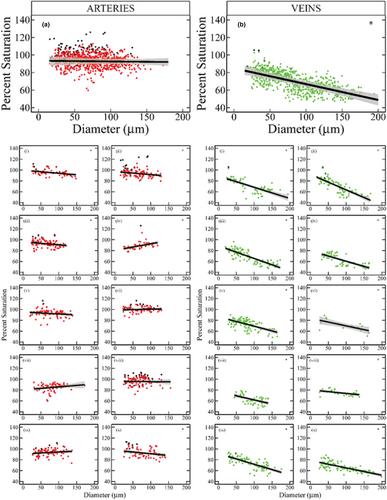
Venous saturation shows an inverse relationship with vessel diameter (D) across subjects, with a mean slope of −0.180 (SE = 0.022) %/μm (20 < D < 180 μm) and a more rapid saturation increase at small vessel diameters reaching to over 80%. Arterial saturation yields smaller positive and negative slopes in individual subjects, with an average of −0.007 (SE = 0.021) %/μm (20 < D < 200 μm) across all subjects. Measurements where vessel brightness exceeds that of the retinal background result in negative values of optical density, causing an artifactual increase in saturation. Optimization of model reflectance values produces a good fit of the conceptual model to measured ODRs.
Conclusion
Measurement artefacts in retinal oximetry are caused by strong central vessel reflections, and apparent diameter sensitivity may result from single and double-pass transmission in vessels. Improvement in correction for vessel diameter is indicated for arteries however further study is necessary for venous corrections.


 求助内容:
求助内容: 应助结果提醒方式:


