Lorenzo Grassi, Sami P. V??n?nen, Lars Jehpsson, ?sten Ljunggren, Bj?rn E. Rosengren, Magnus K. Karlsson, Hanna Isaksson
下载PDF
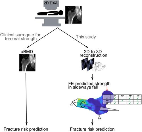
{"title":"3D Finite Element Models Reconstructed From 2D Dual-Energy X-Ray Absorptiometry (DXA) Images Improve Hip Fracture Prediction Compared to Areal BMD in Osteoporotic Fractures in Men (MrOS) Sweden Cohort","authors":"Lorenzo Grassi, Sami P. V??n?nen, Lars Jehpsson, ?sten Ljunggren, Bj?rn E. Rosengren, Magnus K. Karlsson, Hanna Isaksson","doi":"10.1002/jbmr.4878","DOIUrl":null,"url":null,"abstract":"<p>Bone strength is an important contributor to fracture risk. Areal bone mineral density (aBMD) derived from dual-energy X-ray absorptiometry (DXA) is used as a surrogate for bone strength in fracture risk prediction tools. 3D finite element (FE) models predict bone strength better than aBMD, but their clinical use is limited by the need for 3D computed tomography and lack of automation. We have earlier developed a method to reconstruct the 3D hip anatomy from a 2D DXA image, followed by subject-specific FE-based prediction of proximal femoral strength. In the current study, we aim to evaluate the method's ability to predict incident hip fractures in a population-based cohort (Osteoporotic Fractures in Men [MrOS] Sweden). We defined two subcohorts: (i) hip fracture cases and controls cohort: 120 men with a hip fracture (<10 years from baseline) and two controls to each hip fracture case, matched by age, height, and body mass index; and (ii) fallers cohort: 86 men who had fallen the year before their hip DXA scan was acquired, 15 of which sustained a hip fracture during the following 10 years. For each participant, we reconstructed the 3D hip anatomy and predicted proximal femoral strength in 10 sideways fall configurations using FE analysis. The FE-predicted proximal femoral strength was a better predictor of incident hip fractures than aBMD for both hip fracture cases and controls (difference in area under the receiver operating characteristics curve, ΔAUROC = 0.06) and fallers (ΔAUROC = 0.22) cohorts. This is the first time that FE models outperformed aBMD in predicting incident hip fractures in a population-based prospectively followed cohort based on 3D FE models obtained from a 2D DXA scan. Our approach has potential to notably improve the accuracy of fracture risk predictions in a clinically feasible manner (only one single DXA image is needed) and without additional costs compared to the current clinical approach. © 2023 The Authors. <i>Journal of Bone and Mineral Research</i> published by Wiley Periodicals LLC on behalf of American Society for Bone and Mineral Research (ASBMR).</p>","PeriodicalId":185,"journal":{"name":"Journal of Bone and Mineral Research","volume":"38 9","pages":"1258-1267"},"PeriodicalIF":5.1000,"publicationDate":"2023-07-07","publicationTypes":"Journal Article","fieldsOfStudy":null,"isOpenAccess":false,"openAccessPdf":"https://onlinelibrary.wiley.com/doi/epdf/10.1002/jbmr.4878","citationCount":"0","resultStr":null,"platform":"Semanticscholar","paperid":null,"PeriodicalName":"Journal of Bone and Mineral Research","FirstCategoryId":"3","ListUrlMain":"https://onlinelibrary.wiley.com/doi/10.1002/jbmr.4878","RegionNum":1,"RegionCategory":"医学","ArticlePicture":[],"TitleCN":null,"AbstractTextCN":null,"PMCID":null,"EPubDate":"","PubModel":"","JCR":"Q1","JCRName":"ENDOCRINOLOGY & METABOLISM","Score":null,"Total":0}
引用次数: 0
引用
批量引用
Abstract
Bone strength is an important contributor to fracture risk. Areal bone mineral density (aBMD) derived from dual-energy X-ray absorptiometry (DXA) is used as a surrogate for bone strength in fracture risk prediction tools. 3D finite element (FE) models predict bone strength better than aBMD, but their clinical use is limited by the need for 3D computed tomography and lack of automation. We have earlier developed a method to reconstruct the 3D hip anatomy from a 2D DXA image, followed by subject-specific FE-based prediction of proximal femoral strength. In the current study, we aim to evaluate the method's ability to predict incident hip fractures in a population-based cohort (Osteoporotic Fractures in Men [MrOS] Sweden). We defined two subcohorts: (i) hip fracture cases and controls cohort: 120 men with a hip fracture (<10 years from baseline) and two controls to each hip fracture case, matched by age, height, and body mass index; and (ii) fallers cohort: 86 men who had fallen the year before their hip DXA scan was acquired, 15 of which sustained a hip fracture during the following 10 years. For each participant, we reconstructed the 3D hip anatomy and predicted proximal femoral strength in 10 sideways fall configurations using FE analysis. The FE-predicted proximal femoral strength was a better predictor of incident hip fractures than aBMD for both hip fracture cases and controls (difference in area under the receiver operating characteristics curve, ΔAUROC = 0.06) and fallers (ΔAUROC = 0.22) cohorts. This is the first time that FE models outperformed aBMD in predicting incident hip fractures in a population-based prospectively followed cohort based on 3D FE models obtained from a 2D DXA scan. Our approach has potential to notably improve the accuracy of fracture risk predictions in a clinically feasible manner (only one single DXA image is needed) and without additional costs compared to the current clinical approach. © 2023 The Authors. Journal of Bone and Mineral Research published by Wiley Periodicals LLC on behalf of American Society for Bone and Mineral Research (ASBMR).
根据2D双能X射线吸收法(DXA)图像重建的三维有限元模型与瑞典男性骨质疏松性骨折患者区域BMD相比,改善了髋部骨折预测
骨强度是导致骨折风险的重要因素。在骨折风险预测工具中,由双能X射线吸收仪(DXA)得出的区域骨密度(aBMD)被用作骨强度的替代品。三维有限元(FE)模型比aBMD更好地预测骨强度,但由于需要三维计算机断层扫描和缺乏自动化,其临床应用受到限制。我们早些时候开发了一种方法,从2D DXA图像重建3D髋关节解剖结构,然后根据受试者的具体FE预测股骨近端强度。在目前的研究中,我们旨在评估该方法在基于人群的队列中预测髋关节骨折事件的能力(瑞典男性骨质疏松性骨折[MrOS])。我们定义了两个子组:(i)髋部骨折病例和对照组:120名髋部骨折男性(<;10 从基线开始的年数)和两个对照组,根据年龄、身高和体重指数进行匹配;和(ii)跌倒者队列:86名男性在髋关节DXA扫描前一年跌倒,其中15人在接下来的10年中髋关节骨折 年。对于每个参与者,我们重建了三维髋关节解剖结构,并使用有限元分析预测了10种侧向跌倒形态下的股骨近端强度。对于髋部骨折病例和对照组,FE预测的股骨近端强度比aBMD更能预测髋部骨折的发生(受试者操作特征曲线下面积的差异ΔAUROC = 0.06)和下降(ΔAUROC = 0.22)队列。这是首次在基于2D DXA扫描获得的3D FE模型的基于人群的前瞻性随访队列中,FE模型在预测髋关节骨折事件方面优于aBMD。与目前的临床方法相比,我们的方法有可能以临床可行的方式(只需要一张DXA图像)显著提高骨折风险预测的准确性,并且不需要额外的成本。©2023作者。由Wiley Periodicals LLC代表美国骨与矿物研究学会(ASBMR)出版的《骨与矿产研究杂志》。
本文章由计算机程序翻译,如有差异,请以英文原文为准。


 求助内容:
求助内容: 应助结果提醒方式:


