Acute Hydrops with Total Corneal Edema in a Very Young Child with Keratoconus: The Youngest Age Reported Case.
IF 0.4
Q4 OPHTHALMOLOGY
Case Reports in Ophthalmological Medicine
Pub Date : 2022-08-12
eCollection Date: 2022-01-01
DOI:10.1155/2022/2381703
引用次数: 0
Abstract
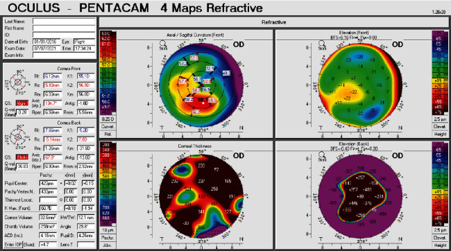
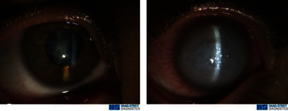
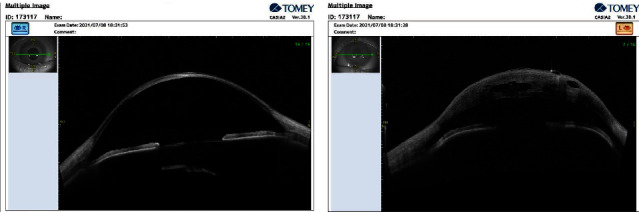
Purpose To present the youngest age ever reported for acute corneal hydrops with total corneal edema in a child with advanced bilateral keratoconus. Methods Patient presentation in ophthalmic clinic. The patient underwent various clinical tests and examinations including anterior segment optical coherence tomography (AS-OCT) and Scheimpflug corneal tomography. Results A 5-year-old girl presented with uncorrected distance visual acuity (UDVA) of 0.4 in the right eye and nonmeasurable UDVA associated with severe photophobia in her left eye of a 3-day duration. Intraocular pressure using the iCare tonometer was 14 and 5 mmHg in the right and left eyes, respectively. An old corneal hydrops scar and posterior subcapsular cataract (PSC) in the right eye and a total limbus to limbus corneal hydrops in the left eye were observed on slit-lamp examinations. Scheimpflug corneal tomography was possible in the right eye but, due to excessive irregularity and scaring, was not possible in the left eye. Corneal thinning and scarring were evident in the anterior segment optical coherence tomography in the right eye and very edematous cornea associated with stromal cleft and epithelial bullae in the left eye. A management plan consisting of topical hypertonic solution and ointment was started to reduce her symptoms. Conclusion Acute corneal hydrops may be the presenting sign of keratoconus; however, extensive hydrops involving the total cornea area at a very young age is very rare and has not been previously reported in the literature.
急性积液伴全角膜水肿的幼童圆锥角膜:最小年龄报告病例。
目的:介绍一名患有晚期双侧圆锥角膜的儿童急性角膜积液合并全角膜水肿的最小年龄。方法:对眼科门诊患者的临床表现进行分析。患者接受了各种临床检查和检查,包括前段光学相干断层扫描(AS-OCT)和Scheimpflug角膜断层扫描。结果:1例5岁女童,右眼未矫正距离视力(UDVA)为0.4,左眼严重畏光,UDVA不可测量,持续3天。使用iCare眼压计,右眼和左眼眼压分别为14和5 mmHg。裂隙灯检查发现右眼陈旧性角膜积液瘢痕及后囊膜下白内障(PSC),左眼角膜缘至角膜缘全积液。在右眼可以进行Scheimpflug角膜断层扫描,但由于过度不规则和惊吓,不可能在左眼进行。右眼前段光学相干断层扫描显示角膜变薄和瘢痕明显,左眼角膜水肿严重,伴有间质裂隙和上皮大泡。治疗方案包括局部高渗溶液和软膏,以减轻她的症状。结论:急性角膜积液可能是圆锥角膜的表现;然而,广泛的积液累及整个角膜区域在非常年轻的年龄是非常罕见的,并没有在以前的文献报道。
本文章由计算机程序翻译,如有差异,请以英文原文为准。
求助全文
约1分钟内获得全文
求助全文

 求助内容:
求助内容: 应助结果提醒方式:


