{"title":"Temporoparietal Brain Hydatid Cyst in an Eight-Year-Old Child: A Rare Case Report.","authors":"Gashaw Arega, Gelassa Merga, Getu Tafa, Fathia Omer Salah, Getnet Abebe, Seblewengel Maru, Wondwossen Ergete","doi":"10.2147/PHMT.S390336","DOIUrl":null,"url":null,"abstract":"<p><p>Hydatidosis is a parasitic disease caused by <i>Echinococcus granulosus</i>, which affects children in many different parts of the world. It commonly affects the lungs and the liver of the children. Brain hydatidosis is an extremely rare clinical condition in the pediatric population, presenting with non-specific symptoms and signs. The diagnosis of intracranial hydatid cysts can be established by brain magnetic resonance imaging and histopathological examination of the specimen. Here, we report an 8-year-old female child diagnosed with a temporoparietal brain hydatid cyst. Brain magnetic resonance imaging showed a thin-walled cystic lesion located in the right temporoparietal lobe with significant mass effect and midline shift, with no abnormal wall or solid enhancement, and no surrounding edema. The diagnosis of brain temporoparietal hydatid cyst was made radiologically. The patient was operated on and the cyst was completely removed without rupture. The removed cyst was sent for histopathological examination; the histological sections showed a laminated acellular cyst wall with a nucleated germinal layer and no protoscolices, and the diagnosis of temporoparietal brain hydatid cyst was confirmed. The patient had a smooth postoperative course, started albendazole therapy, and was discharged with improvement.</p>","PeriodicalId":74410,"journal":{"name":"Pediatric health, medicine and therapeutics","volume":" ","pages":"361-365"},"PeriodicalIF":1.7000,"publicationDate":"2022-11-10","publicationTypes":"Journal Article","fieldsOfStudy":null,"isOpenAccess":false,"openAccessPdf":"https://ftp.ncbi.nlm.nih.gov/pub/pmc/oa_pdf/4a/5a/phmt-13-361.PMC9662011.pdf","citationCount":"1","resultStr":null,"platform":"Semanticscholar","paperid":null,"PeriodicalName":"Pediatric health, medicine and therapeutics","FirstCategoryId":"1085","ListUrlMain":"https://doi.org/10.2147/PHMT.S390336","RegionNum":0,"RegionCategory":null,"ArticlePicture":[],"TitleCN":null,"AbstractTextCN":null,"PMCID":null,"EPubDate":"2022/1/1 0:00:00","PubModel":"eCollection","JCR":"Q2","JCRName":"PEDIATRICS","Score":null,"Total":0}
引用次数: 1
Abstract
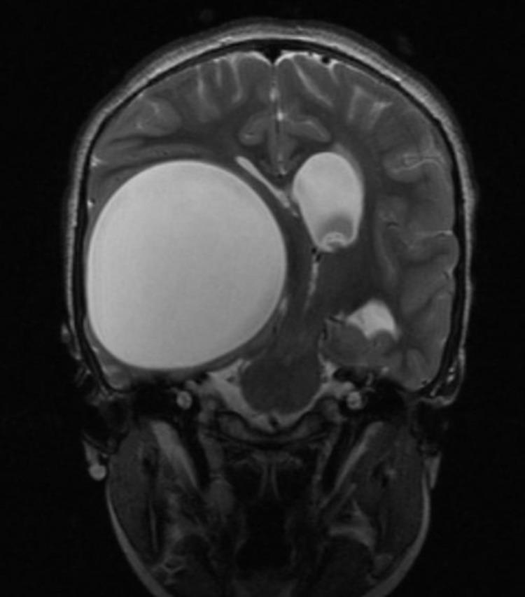
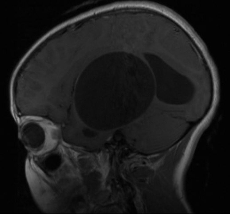
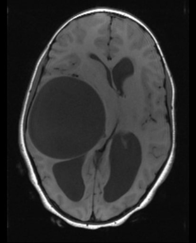
Hydatidosis is a parasitic disease caused by Echinococcus granulosus, which affects children in many different parts of the world. It commonly affects the lungs and the liver of the children. Brain hydatidosis is an extremely rare clinical condition in the pediatric population, presenting with non-specific symptoms and signs. The diagnosis of intracranial hydatid cysts can be established by brain magnetic resonance imaging and histopathological examination of the specimen. Here, we report an 8-year-old female child diagnosed with a temporoparietal brain hydatid cyst. Brain magnetic resonance imaging showed a thin-walled cystic lesion located in the right temporoparietal lobe with significant mass effect and midline shift, with no abnormal wall or solid enhancement, and no surrounding edema. The diagnosis of brain temporoparietal hydatid cyst was made radiologically. The patient was operated on and the cyst was completely removed without rupture. The removed cyst was sent for histopathological examination; the histological sections showed a laminated acellular cyst wall with a nucleated germinal layer and no protoscolices, and the diagnosis of temporoparietal brain hydatid cyst was confirmed. The patient had a smooth postoperative course, started albendazole therapy, and was discharged with improvement.

 求助内容:
求助内容: 应助结果提醒方式:


