Comparative evaluation of the effects of three hydraulic calcium silicate cements on odontoblastic differentiation of human dental pulp stem cells: an in vitro study.
Hadi Assadian, Arash Khojasteh, Zahra Ebrahimian, Fereshteh Ahmadinejad, Helia Sadat Haeri Boroojeni, Mahboubeh Bohlouli, Mohammad Hossein Nekoofar, Paul Mh Dummer, Hanieh Nokhbatolfoghahaei
{"title":"Comparative evaluation of the effects of three hydraulic calcium silicate cements on odontoblastic differentiation of human dental pulp stem cells: an in vitro study.","authors":"Hadi Assadian, Arash Khojasteh, Zahra Ebrahimian, Fereshteh Ahmadinejad, Helia Sadat Haeri Boroojeni, Mahboubeh Bohlouli, Mohammad Hossein Nekoofar, Paul Mh Dummer, Hanieh Nokhbatolfoghahaei","doi":"10.1590/1678-7757-2022-0203","DOIUrl":null,"url":null,"abstract":"<p><strong>Objective: </strong>The study aimed to compare the response of human dental pulp stem cells (hDPSCs) towards three hydraulic calcium silicate cements (HCSCs) by measuring cytotoxicity and expression of dentinogenic genes.</p><p><strong>Methodology: </strong>Dental pulps of five impacted mandibular third molars were extirpated as a source for hDPSCs. Next to culturing, hDPSCs were subjected to fluorescence-activated cell sorting after the third passage to validate stemness of the cells. Human DPSCs were exposed to diluted supernatants of OrthoMTA (OMTA), Biodentine (BD) and Calcium-Enriched Mixture (CEM) at concentrations 10, 25, 50 and 100% at the first, third and fifth day of culture. Then, cells were exposed to 10% concentrations supernatant of HCSCs to determine DSPP and DMP1 gene expression, using a quantitative polymerase-chain reaction. Data were analyzed using one-way and three-way ANOVA, followed by Tukey post hoc statistical tests.</p><p><strong>Results: </strong>Optimal cell proliferation was observed in all groups, regardless of concentration and time-point. HCSC supernatants were non-cytotoxic to hDPSCs at all three time-points, except for 100% Biodentine on day five. On day seven, OMTA group significantly upregulated the expression of DSPP and DMP1 genes. On day 14, expression of DMP1 and DSPP genes were significantly higher in BD and OMTA groups, respectively.</p><p><strong>Conclusion: </strong>Biodentine significantly upregulated DMP1 gene expression over 14 days, whereas CEM was associated with only minimal expression of DSPP and DMP1 .</p>","PeriodicalId":321675,"journal":{"name":"Journal of applied oral science : revista FOB","volume":" ","pages":"e20220203"},"PeriodicalIF":0.0000,"publicationDate":"2022-11-04","publicationTypes":"Journal Article","fieldsOfStudy":null,"isOpenAccess":false,"openAccessPdf":"https://www.ncbi.nlm.nih.gov/pmc/articles/PMC9651926/pdf/","citationCount":"1","resultStr":null,"platform":"Semanticscholar","paperid":null,"PeriodicalName":"Journal of applied oral science : revista FOB","FirstCategoryId":"3","ListUrlMain":"https://doi.org/10.1590/1678-7757-2022-0203","RegionNum":0,"RegionCategory":null,"ArticlePicture":[],"TitleCN":null,"AbstractTextCN":null,"PMCID":null,"EPubDate":"2022/1/1 0:00:00","PubModel":"eCollection","JCR":"","JCRName":"","Score":null,"Total":0}
引用次数: 1
Abstract
Objective: The study aimed to compare the response of human dental pulp stem cells (hDPSCs) towards three hydraulic calcium silicate cements (HCSCs) by measuring cytotoxicity and expression of dentinogenic genes.
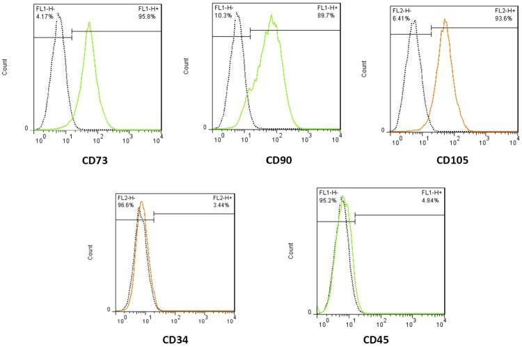
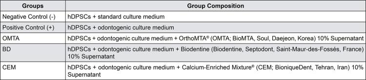
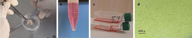
Methodology: Dental pulps of five impacted mandibular third molars were extirpated as a source for hDPSCs. Next to culturing, hDPSCs were subjected to fluorescence-activated cell sorting after the third passage to validate stemness of the cells. Human DPSCs were exposed to diluted supernatants of OrthoMTA (OMTA), Biodentine (BD) and Calcium-Enriched Mixture (CEM) at concentrations 10, 25, 50 and 100% at the first, third and fifth day of culture. Then, cells were exposed to 10% concentrations supernatant of HCSCs to determine DSPP and DMP1 gene expression, using a quantitative polymerase-chain reaction. Data were analyzed using one-way and three-way ANOVA, followed by Tukey post hoc statistical tests.
Results: Optimal cell proliferation was observed in all groups, regardless of concentration and time-point. HCSC supernatants were non-cytotoxic to hDPSCs at all three time-points, except for 100% Biodentine on day five. On day seven, OMTA group significantly upregulated the expression of DSPP and DMP1 genes. On day 14, expression of DMP1 and DSPP genes were significantly higher in BD and OMTA groups, respectively.
Conclusion: Biodentine significantly upregulated DMP1 gene expression over 14 days, whereas CEM was associated with only minimal expression of DSPP and DMP1 .

 求助内容:
求助内容: 应助结果提醒方式:


