Maria Lage Barca, Dag Alnæs, Knut Engedal, Karin Persson, Rannveig Sakshaug Eldholm, Nikias Siafarikas, Ina Selseth Almdahl, Maria Stylianou-Korsnes, Ingvild Saltvedt, Geir Selbæk, Lars T Westlye
{"title":"Brain Morphometric Correlates of Depressive Symptoms among Patients with and without Dementia.","authors":"Maria Lage Barca, Dag Alnæs, Knut Engedal, Karin Persson, Rannveig Sakshaug Eldholm, Nikias Siafarikas, Ina Selseth Almdahl, Maria Stylianou-Korsnes, Ingvild Saltvedt, Geir Selbæk, Lars T Westlye","doi":"10.1159/000521114","DOIUrl":null,"url":null,"abstract":"<p><strong>Introduction: </strong>Findings regarding brain morphometry among patients with dementia and concomitant depressive symptoms have been inconsistent. Thus, the aim of the present study was to test the hypothesis that dementia and concomitant depressive symptoms are associated with structural brain changes in the temporal lobe measured with structural magnetic resonance imaging (MRI).</p><p><strong>Methods: </strong>A sample of 492 patients from Norwegian memory clinics (<i>n</i> = 363) and Old Age Psychiatry services (<i>n</i> = 129) was studied. The assessment included the Cornell Scale for Depression in Dementia (CSDD), Instrumental Activities of Daily Living Scale, Mini Mental State Examination, and MRI of the brain, processed with FreeSurfer to derive ROI measures of cortical thickness, volume, and area using the Desikan-Killiany parcellation, as well as subcortical volumes. Dementia was diagnosed according to ICD-10 research criteria. Correlates of brain morphometry using multiple linear regression were examined.</p><p><strong>Results: </strong>Higher scores on the CSDD were associated with larger cortical volume (β = 0.125; <i>p</i> value = 0.003) and area of the left isthmus of the cingulate gyrus (β = 0.151; <i>p</i> value = <0.001) across all patients. Inclusion of an interaction term (dementia × CSDD) revealed a smaller area in the left temporal pole (β = -0.345; <i>p</i> value = 0.001) and right-transverse temporal cortex (β = -0.321; <i>p</i> value = 0.001) in patients with dementia and depressive symptoms.</p><p><strong>Discussion/conclusion: </strong>We confirm the previous findings of structural brain changes in temporal regions among patients with dementia and concomitant depressive symptoms. This may contribute to a better understanding of the mechanisms underlying depression in dementia. To the best of our knowledge, this is the largest study conducted on this topic to date.</p>","PeriodicalId":38017,"journal":{"name":"Dementia and Geriatric Cognitive Disorders Extra","volume":" ","pages":"107-114"},"PeriodicalIF":1.6000,"publicationDate":"2022-06-23","publicationTypes":"Journal Article","fieldsOfStudy":null,"isOpenAccess":false,"openAccessPdf":"https://ftp.ncbi.nlm.nih.gov/pub/pmc/oa_pdf/ed/58/dee-0012-0107.PMC9251457.pdf","citationCount":"1","resultStr":null,"platform":"Semanticscholar","paperid":null,"PeriodicalName":"Dementia and Geriatric Cognitive Disorders Extra","FirstCategoryId":"1085","ListUrlMain":"https://doi.org/10.1159/000521114","RegionNum":0,"RegionCategory":null,"ArticlePicture":[],"TitleCN":null,"AbstractTextCN":null,"PMCID":null,"EPubDate":"2022/5/1 0:00:00","PubModel":"eCollection","JCR":"Q4","JCRName":"CLINICAL NEUROLOGY","Score":null,"Total":0}
引用次数: 1
Abstract
Introduction: Findings regarding brain morphometry among patients with dementia and concomitant depressive symptoms have been inconsistent. Thus, the aim of the present study was to test the hypothesis that dementia and concomitant depressive symptoms are associated with structural brain changes in the temporal lobe measured with structural magnetic resonance imaging (MRI).
Methods: A sample of 492 patients from Norwegian memory clinics (n = 363) and Old Age Psychiatry services (n = 129) was studied. The assessment included the Cornell Scale for Depression in Dementia (CSDD), Instrumental Activities of Daily Living Scale, Mini Mental State Examination, and MRI of the brain, processed with FreeSurfer to derive ROI measures of cortical thickness, volume, and area using the Desikan-Killiany parcellation, as well as subcortical volumes. Dementia was diagnosed according to ICD-10 research criteria. Correlates of brain morphometry using multiple linear regression were examined.
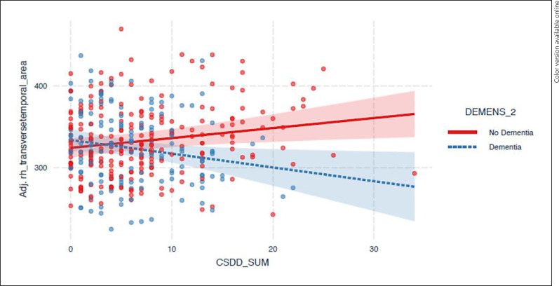
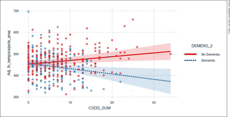
Results: Higher scores on the CSDD were associated with larger cortical volume (β = 0.125; p value = 0.003) and area of the left isthmus of the cingulate gyrus (β = 0.151; p value = <0.001) across all patients. Inclusion of an interaction term (dementia × CSDD) revealed a smaller area in the left temporal pole (β = -0.345; p value = 0.001) and right-transverse temporal cortex (β = -0.321; p value = 0.001) in patients with dementia and depressive symptoms.
Discussion/conclusion: We confirm the previous findings of structural brain changes in temporal regions among patients with dementia and concomitant depressive symptoms. This may contribute to a better understanding of the mechanisms underlying depression in dementia. To the best of our knowledge, this is the largest study conducted on this topic to date.
期刊介绍:
This open access and online-only journal publishes original articles covering the entire spectrum of cognitive dysfunction such as Alzheimer’s and Parkinson’s disease, Huntington’s chorea and other neurodegenerative diseases. The journal draws from diverse related research disciplines such as psychogeriatrics, neuropsychology, clinical neurology, morphology, physiology, genetic molecular biology, pathology, biochemistry, immunology, pharmacology and pharmaceutics. Strong emphasis is placed on the publication of research findings from animal studies which are complemented by clinical and therapeutic experience to give an overall appreciation of the field. Dementia and Geriatric Cognitive Disorders Extra provides additional contents based on reviewed and accepted submissions to the main journal Dementia and Geriatric Cognitive Disorders Extra .

 求助内容:
求助内容: 应助结果提醒方式:


