Jannik von Schöning, Jörg Flitsch, Dieter K Lüdecke, Rudolf Fahlbusch, Michael Buchfelder, Rolf Buslei, Ulrich J Knappe, Markus Bergmann, Walter J Schulz-Schaeffer, Jochen Herms, Markus Glatzel, Wolfgang Saeger
{"title":"Multiple tumorous lesions of the pituitary gland.","authors":"Jannik von Schöning, Jörg Flitsch, Dieter K Lüdecke, Rudolf Fahlbusch, Michael Buchfelder, Rolf Buslei, Ulrich J Knappe, Markus Bergmann, Walter J Schulz-Schaeffer, Jochen Herms, Markus Glatzel, Wolfgang Saeger","doi":"10.1007/s42000-022-00392-9","DOIUrl":null,"url":null,"abstract":"<p><strong>Purpose/objective: </strong>Multiple tumorous lesions in one pituitary gland are rare and mostly described in case reports. Their incidences and combinations are defined in larger collectives. Therefore, we analyzed our large collection for double tumors and combinations of tumors, cysts, and inflammation.</p><p><strong>Methods: </strong>The German Registry of Pituitary Tumors, including cases from 1990 to 2018, served as the database. Our collection comprises a total of 16,283 cases up until the end of 2018. Of these cases, 12,673 originated from surgical and 3,610 from autopsy material. All specimens were fixed in formalin and embedded in paraffin. The sections were stained with hematoxylin-eosin and PAS. Monoclonal (prolactin, TSH, FSH, LH, and α subunit) or polyclonal (GH and ACTH) antibodies were used to detect pituitary hormones in the lesions. Since 2017, antibodies against the transcription factors Pit-1, T-Pit, and SF-1 have been used in difficult cases. The criteria of the 2017 WHO classification have been basic principles for classification since 2018 (Osamura et al. 2017). For differentiation of other sellar tumors, such as meningiomas, chordomas, or metastases, the use of additional antibodies was necessary. For these cases, it was possible to use a broad antibody spectrum. Autopsy pituitaries were generally studied by H&E and PAS sections. If any lesions were demonstrated in these specimens, additional immunostaining was performed.</p><p><strong>Results: </strong>Multiple tumorous lesions with more than one pituitary neuroendocrine tumor (PitNET) respectively adenoma make up 1.4% (232 cases) in our collection. Within the selected cases, synchronous multiple pituitary neuroendocrine tumors (PitNETs) account for 17.3%, PANCH cases (pituitary adenoma with neuronal choristoma) for 14.7%, PitNETs and posterior lobe tumors for 2.2%, PitNETs and metastases for 5.2%, PitNETs and mesenchymal tumors for 2.6%, PitNETs and cysts for 52.2%, and PitNETs and primary inflammation for 6.0%. The mean patient age was 53.8 years, with a standard deviation of 18.5 years. A total of 55.3% of the patients were female and 44.7% were male. From 1990 to 2018, there was a continuous increase in the number of multiple tumorous lesions.</p><p><strong>Conclusion: </strong>From our studies, we conclude that considering possible tumorous double lesions during surgeries and in preoperative X-ray analyses is recommended.</p>","PeriodicalId":520640,"journal":{"name":"Hormones (Athens, Greece)","volume":" ","pages":"653-663"},"PeriodicalIF":0.0000,"publicationDate":"2022-12-01","publicationTypes":"Journal Article","fieldsOfStudy":null,"isOpenAccess":false,"openAccessPdf":"https://www.ncbi.nlm.nih.gov/pmc/articles/PMC9712358/pdf/","citationCount":"3","resultStr":null,"platform":"Semanticscholar","paperid":null,"PeriodicalName":"Hormones (Athens, Greece)","FirstCategoryId":"3","ListUrlMain":"https://doi.org/10.1007/s42000-022-00392-9","RegionNum":0,"RegionCategory":null,"ArticlePicture":[],"TitleCN":null,"AbstractTextCN":null,"PMCID":null,"EPubDate":"2022/8/10 0:00:00","PubModel":"Epub","JCR":"","JCRName":"","Score":null,"Total":0}
引用次数: 3
Abstract
Purpose/objective: Multiple tumorous lesions in one pituitary gland are rare and mostly described in case reports. Their incidences and combinations are defined in larger collectives. Therefore, we analyzed our large collection for double tumors and combinations of tumors, cysts, and inflammation.
Methods: The German Registry of Pituitary Tumors, including cases from 1990 to 2018, served as the database. Our collection comprises a total of 16,283 cases up until the end of 2018. Of these cases, 12,673 originated from surgical and 3,610 from autopsy material. All specimens were fixed in formalin and embedded in paraffin. The sections were stained with hematoxylin-eosin and PAS. Monoclonal (prolactin, TSH, FSH, LH, and α subunit) or polyclonal (GH and ACTH) antibodies were used to detect pituitary hormones in the lesions. Since 2017, antibodies against the transcription factors Pit-1, T-Pit, and SF-1 have been used in difficult cases. The criteria of the 2017 WHO classification have been basic principles for classification since 2018 (Osamura et al. 2017). For differentiation of other sellar tumors, such as meningiomas, chordomas, or metastases, the use of additional antibodies was necessary. For these cases, it was possible to use a broad antibody spectrum. Autopsy pituitaries were generally studied by H&E and PAS sections. If any lesions were demonstrated in these specimens, additional immunostaining was performed.
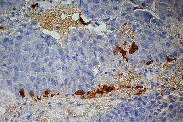
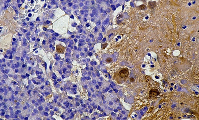
Results: Multiple tumorous lesions with more than one pituitary neuroendocrine tumor (PitNET) respectively adenoma make up 1.4% (232 cases) in our collection. Within the selected cases, synchronous multiple pituitary neuroendocrine tumors (PitNETs) account for 17.3%, PANCH cases (pituitary adenoma with neuronal choristoma) for 14.7%, PitNETs and posterior lobe tumors for 2.2%, PitNETs and metastases for 5.2%, PitNETs and mesenchymal tumors for 2.6%, PitNETs and cysts for 52.2%, and PitNETs and primary inflammation for 6.0%. The mean patient age was 53.8 years, with a standard deviation of 18.5 years. A total of 55.3% of the patients were female and 44.7% were male. From 1990 to 2018, there was a continuous increase in the number of multiple tumorous lesions.
Conclusion: From our studies, we conclude that considering possible tumorous double lesions during surgeries and in preoperative X-ray analyses is recommended.
目的/目的:在一个垂体多发肿瘤病变是罕见的,大多在病例报告中描述。它们的发生率和组合在更大的集体中定义。因此,我们分析了我们收集的大量双重肿瘤和肿瘤、囊肿和炎症的组合。方法:以德国垂体肿瘤登记处(German Registry of Pituitary Tumors)为数据库,包括1990年至2018年的病例。截至2018年底,我们的收藏共计16283件。在这些病例中,12,673例来自外科手术,3,610例来自尸检材料。所有标本用福尔马林固定,石蜡包埋。切片用苏木精-伊红和PAS染色。采用单克隆抗体(催乳素、TSH、FSH、LH和α亚基)或多克隆抗体(GH和ACTH)检测病灶内垂体激素。自2017年以来,针对转录因子Pit-1、T-Pit和SF-1的抗体已用于治疗困难病例。自2018年以来,2017年世卫组织分类标准一直是分类的基本原则(Osamura et al. 2017)。对于其他鞍区肿瘤的分化,如脑膜瘤、脊索瘤或转移瘤,需要使用额外的抗体。对于这些病例,可以使用广泛的抗体谱。尸检垂体一般通过H&E和PAS切片进行研究。如果在这些标本中发现任何病变,则进行额外的免疫染色。结果:本组病例中,合并一个以上垂体神经内分泌肿瘤(PitNET)的多发肿瘤占1.4%(232例)。在所选病例中,同步多发性垂体神经内分泌肿瘤(PitNETs)占17.3%,PANCH(垂体腺瘤合并神经毛膜瘤)占14.7%,PitNETs合并后叶肿瘤占2.2%,PitNETs合并转移瘤占5.2%,PitNETs合并间质肿瘤占2.6%,PitNETs合并囊肿占52.2%,PitNETs合并原发性炎症占6.0%。患者平均年龄为53.8岁,标准差为18.5岁。女性占55.3%,男性占44.7%。从1990年到2018年,多发性肿瘤病变数量持续增加。结论:从我们的研究中,我们得出结论,建议在手术和术前x线分析中考虑可能的肿瘤双重病变。

 求助内容:
求助内容: 应助结果提醒方式:


